16 June, 2013

Japanese Scientists Debunk Longstanding Avian Behavioral Theory

Recent research published in the Journal Amyloid casts doubt on the widely accepted concept that chickens cross the road merely to get to the other side. It appears that at least some of them may actually be searching for an amyloidosis specialist. 

Tomoaki Murakami and colleagues describe their research related to an outbreak of AA amyloidosis occurring at a Japanese poultry farm. Avian AA amyloidosis is well characterized (as indicated by its inclusion in Table II from the most recent amyloid fibril nomenclature committee guidelines). AA amyloidosis has been described in waterfowl (due to chronic inflammatory conditions) and chickens (due to bacterial infections or repeated vaccinations). AA amyloid has also been detected in the livers of force-fed ducks and geese used for foie gras. 

A photomicrograph of a hepatic venule from a sample of commercially-available foie gras
(top panel, h/e stain; middle, congo red stain under polarized light; bottom IHC confirming AA amyloid).
Ref: http://www.ncbi.nlm.nih.gov/pubmed/17578924
The authors studied four breeds of chickens, one of which has previously been shown to have a propensity for developing systemic AA amyloidosis after repeated vaccinations for Salmonella enteritidis (SE) and/or Mycoplasma gallisepticum (MG). In this study, chickens were inoculated with a single dose of one of two SE vaccines ("SE(a)" or "SE(b)") or an MG vaccine. Then some of the chickens were also given a dose of an AA amyloid fibril solution which the authors had prepared from the livers of other chickens with AA amyloidosis. Additionally, there were some chickens who received only vaccine without AA fibrils, only fibrils without vaccine, or neither.

The findings: 

  • 29 out of 38 chickens (all breeds studied) who got both vaccine SE(a) and a dose of AA fibrils (either orally or intravenously) developed AA amyloidosis. If the AA fibrils were administered orally, then the amyloidosis tended to develop in the chickens' spleens; the chickens who got the AA fibrils intravenously developed more widespread systemic amyloidosis (including the birds' intestines). 
  • ZERO out of 113 other chickens developed systemic AA amyloidosis.
  • Serum AA precursor protein levels (SAA levels) did not correlate with risk of developing systemic AA amyloidosis. The SE(a) vaccine did not induce an increase in SAA in any subgroup of chickens (including those who developed amyloidosis) and the MG vaccine did induce a rise in SAA (with no cases of associated amyloidosis).
The scientists (and another author providing commentary in the same issue of the journal) concluded that the outbreak of avian AA amyloidosis was likely the result of chickens ingesting amyloid-contaminated feed and/or droppings after having had industry-standard vaccinations which may have pre-disposed them to developing amyloidosis. 

Not an unprecedented finding. Fecal transmission is suspected to contribute to the high incidence of systemic AA amyloidosis in captive cheetahs (like cheetahs don't have enough problems). Also, the same scientists who discovered amyloidosis in foie gras fed it to amyloid-susceptible mice and found it accelerated the rate at which systemic AA amyloidosis developed. They concluded foie gras was a potential Amyloid Enhancing Factor (AEF), and that 
"It would seem prudent for children and adults with rheumatoid arthritis or other diseases who are at risk for [AA amyloidosis] to avoid foods that may be contaminated with AA fibrils." 
Then, citing studies such as one in which formation of a specific type of amyloidosis (AApoAII) could be accelerated in predisposed experimental mice by intravenous injection of any of several different types of human amyloid fibrils (AL, ATTR, Abeta2-MG, and others), they suggest
"...that it may be hazardous for individuals who are prone to develop other types of amyloid-associated disorders, e.g., Alzheimer's disease or type II diabetes, to consume such products."
Which nags at me. I don't so much wonder about whether my father, the son of a woman who died of Alzheimer's disease, should enjoy foie gras as much as he does (though maybe I should - it's Fathers' Day). I worry a little bit about whether diet could be affecting the outcome of my patients with documented systemic amyloidosis. Is our carnivorous American diet full of AEFs? Possibly. An examination of the kidneys and other organs from 302 apparently healthy cattle slaughtered for meat revealed AA amyloidosis in 5% of the animals (take a look). The incidence was lower (0.4% - 2%) in other studies. A largely vegetarian diet has been cited as a possible explanation for the virtual absence of secondary amyloidosis in leprosy patients in India compared to the fairly high incidence amongst leprosy patients in the United States, Malaysia, Brazil, and other countries.  Check out this 1965 article by Amyloid Guru Alan S. Cohen and colleagues which explores this very topic.  At this time, though, there is no compelling evidence for transmission or acceleration of amyloidosis (AA or any other type) in humans from ingestion of AA-containing bovine meat or organs.  Additionally, a search of the medical literature did not turn up any studies looking at the impact of diet on prognosis in AL amyloidosis. So for now, I'll just worry a little bit. Comments welcome. 

19 May, 2013

Digoxin Sensitivity and AL #Amyloidosis

Last week, I learned something. Since learning new things is a good habit to get into, I thought I'd share - especially since the pertinent data were actually a little difficult to track down. 

A patient of mine with long-standing AL amyloidosis was admitted to the hospital with congestive heart failure occurring in the setting of rapid atrial fibrillation ("AFib"). For non-medical folks, this means the top chambers of the heart  (the atria) were beating extremely fast and irregularly, driving the bottom part of the heart (the ventricles) to do the same. This is not a kind thing of the atria to ask of the stiff, thick, amyloid-filled ventricles. 

The situation was particularly difficult because of the patient's chronically low blood pressure.  His typical BP of 90/50 was running even lower due to the abnormal heart rhythm.  Some of the medications one might normally employ to control the Afib - like beta-blockers or calcium channel blockers - could not be used here because of the BP.  I suggested to the medical residents that we try using digoxin, which would not be expected to lower the blood pressure.  An hour later, the team called me back to let me know they had decided against that because the risk of digoxin toxicity was too high in this patient. I asked why they felt he was at risk for this, and the answer surprised me.

"His amyloidosis."

A search of PubMed using the search terms "digoxin" and "amyloidosis" yields only 8 references, at least as of the afternoon of May 18th, 2013. Of these, one of them was a case report of a person who was diagnosed with cardiac amyloidosis after developing fainting spells on digoxin.  Another article describing two cases of familial transthyretin (ATTR) mentions in the abstract that such patients are prone to digoxin toxicity, but does not state that the patients in that report experienced this. I have thus far not been able to track don a copy of the actual article text. The other 6 references that came up were not relevant to my specific patient's case. 

I tried another strategy and pulled up a review from cardiac amyloidosis guru Dr. Rodney Falk. Here is his commentary on the use of digoxin in AL amyloidosis patients: 
"There is no role for digoxin in patients with cardiac amyloidosis who are in sinus rhythm. However, for patients with atrial fibrillation, cautious use of digoxin may aid in heart rate control although the risk of digoxin toxicity may be increased, possibly related to abnormal binding of the drug to amyloid fibrils."
A reference for last statement not provided, so more digging was needed.  Turned up the following additional references, not identified by original search:

A 1961 article from one of Michigan's own in the Annals of Internal Medicine which described two amyloidosis patients who seemed to have problems stemming from digitalis (a drug structurally related to digoxin, but with a longer half-life in the blood: 5-7 days, compared to 1.5-2 days). Pt #1 was a 58-yo farmer with jaundice and hypoalbuminemia (low serum protein levels) and atrial fibrillation who developed severe bradycardia (slow heart rate) of approximately 30 beats per minute after a 0.8 mg loading dose of digitalis followed a couple of days later by a 0.1 mg dose. Although it is not entirely clear from the text, it appears the patient had a liver biopsy proving he had systemic amyloidosis a few days before dying of liver failure.  No information was provided regarding kidney function. On autopsy, multiple organs were confirmed to be infiltrated with amyloid, including the heart. Amyloid fibril typing was not performed.  Pt #2, on the other hand, almost definitely had AL amyloidosis complicating a plasmacytoma. She was in sinus rhythm but had symptoms of congestive heart failure and exam findings suggesting cardiac tamponade (compressive fluid around the heart). She was treated with 1.3 grams of an older digitalis preparation (digitalis folia) over 60 hours. I cannot find a good reference with a dosage equivalency table to put this into current medical context. Also, again, no information on kidney function was provided. After dosing, the patient developed cardiac bigeminy (paired heart beats) with a rate of 70 beats per minute. The patient then developed severe hyperkalemia (high serum potassium levels - 8.3 mEq/L in this case, or about twice the normal level, after originally starting in the normal range). The patient suffered heart rhythm disturbances typical of this potassium level, and she died. On autopsy, the patient was found to have cardiac amyloidosis and a pericardial effusion (fluid around the heart) was confirmed. In 1961, serum digitalis levels were not obtainable.

A second article from A. Pomerance of London's Central Middlesex Hospital's Department of Morbid Anatomy and Histology was received at the British Heart Journal on Aug 17th, 1964, and was ultimately published in 1965. It turns out that at the exact same time, the Beatles were at work trying to get the tracks for Beatles for Sale! recorded and mixed. Busy time for the British. Pomerance's article is an autopsy series of 21 elderly patients with "senile amyloidosis."  At the time, the composition of the fibrils had not been characterized; today we know that most if not all of these cases were likely ATTR (wild type) amyloid.  The report is actually pretty fascinating from a historical standpoint, and it summarizes what was known at the time about systemic amyloidosis citing even older literature. As far as the cases, 3 of 21 patients were reported to have been "sensitive to digitalis" during hospital admissions prior to the terminal ones. No information about digitalis dosing, serum levels, or kidney function was provided. Two of the three patients had had prior myocardial infarctions. 

So, 6 total cases of amyloidosis patients experiencing "digitalis sensitivity." One of the cases provides sufficient detail to justify this diagnosis.  One (pt #2 from the 1961 Cassidy article) almost certainly did not experience digitalis sensitivity but died of hyperkalemia-induced arrhythmia in the setting of pre-existing cardiac tamponade. There is insufficient data on the other four patients (from Pomerance's report, and from the original PubMed search I did) to reach any conclusions one way or the other. 

What about the last part of the cited text from the Falk article? The best support for this was a brief report by Dr A. Rubinow, Martha Skinner, and Alan Cohen from Boston University published in 1981. They describe a laboratory experiment in which they added digoxin to pellets of amyloid fibrils isolated from the spleens of three patients with amyloidosis, and also to samples of ground up tissues: normal human liver, normal human heart, and human heart affected by amyloidosis.  They demonstrated that both pellets of pure amyloid fibrils or heart tissue from a patient with amyloidotic cardiomyopathy bound digoxin, whereas the control tissues (the ones without amyloid) did not. 


Ref: http://circ.ahajournals.org/content/63/6/1285.long
The authors discuss cardiac glycosides' mechanism of action, and - citing the relevance of cardiac tissue levels of the drugs rather than serum levels - postulate that binding of digoxin by amyloid-laden cardiac tissue may increase effective drug levels where it counts. However, they (correctly) point out that it is unknown whether amyloid-bound digoxin retains its pharmacologic activity. Their conclusions? 
"Therapeutic judgement regarding the cautious administration of digoxin in patients with cardiac amyloidosis still rests on clinical grounds."
My conclusions? The same. Patients with amyloid cardiomyopathy are known to be at risk for potentially fatal arrhythmias - its what accounts for the dismal outcomes seen in patients with cardiac stage 3 AL amyloidosis.  It is also clear that patients with structural heart injury from any cause may be at risk for digoxin-induced arrhythmias. So caution is warranted.  A critical review of the literature, however, does not make the case that digoxin can never be used in patients with systemic amyloidosis.  

17 May, 2013

Successful #Amyloidosis Foundation Benefit: Art, Autos, and Amyloid

Tonight, I attended a fine benefit event which raised funds for the Amyloidosis Foundation. I was joined by two other people from the Karmanos Cancer Institute Myeloma and Amyloidosis team (Silva Pregja and Christy Houde). The event was held at the Inn at St. John's in Plymouth, MI. Here is a picture of the room: 


The Atrium Ballroom at the Inn at St. John's. Image downloaded from the I@SJ's website: http://www.stjohnsgolfconference.com/index.cfm

Very nice night with strolling dinner and a silent auction. Over a hundred people in attendance. The event Co-Chair was the daughter of one of my former patients. I had the opportunity to say a few words at the start of the evening describing the good work the Foundation has been doing for the last decade (educating patients and their physicians about amyloidosis - see link to video of GREAT symposium the Karmanos Cancer Institute and Wayne State University co-sponsored with the Amyloidosis Foundation; funding Junior and Senior level research grants to the tune of three-quarters of a million dollars in total).

Also while there, I had the opportunity to speak with some folks from Millennium, Alnylam, and Prothena - All sponsors of the event, and all in the business of developing amyloidosis therapies (Millennium - Bortezomib, Ixazomib; Alnylam - ALN-TTR02, ALN-TTRsc; Prothena - NEOD001).  

As for the auction, I made a successful run at at a nice piece of art:


Aptly named "Horses" (pardon less-than-professional-quality photo).
A nice little pic for the house and a few bucks for a good cause. 


12 May, 2013

Patient Page: Why is AL #amyloidosis bad for kidneys?

"Amyloidosis" refers to any one of several diseases in which abnormal protein fibrils accumulate in a person's organs. The most common type is called AL amyloidosis, and the protein fibrils are made up of fragments of antibody proteins called light chains. I posted a slide which illustrates what a light chain is in a previous post ("Why is AL amyloid bad for hearts?"). In AL amyloidosis, the levels of light chains (usually lambda type, but sometimes kappa type) in the blood and urine are elevated.  Light chains are made by plasma cells in the bone marrow, and current AL amyloid therapy targets these plasma cells. 

Any organ's function can be compromised by amyloid deposits.  The kidney is one of the most commonly affected organs, and patients with injured kidneys may have symptoms, including swelling (edema) of the legs, decreased urine output, and lightheadedness due to sudden drops in blood pressure. 

In order to understand how amyloid injures the kidneys, it is helpful to understand how the kidney works. 

A summary of kidney anatomy and function relevant to amyloidosis:

  • The kidney is made up of a million microscopic filtration units called nephrons. 
  • Each nephron has a filter called the glomerulus, which filters the blood.
  • Some parts of the blood, namely water and electrolytes (sodium, potassium, etc), flow through this filter into a tube system where it is processed further. The material that eventually comes out of the end of all this tubing is urine
  • Other components of the blood, like red blood cells, do not pass through the filter.  
  • Although under normal circumstances there is essentially no protein in our urine, it is not because proteins do not pass through the filter. Filtered protein may be reabsorbed (taken back up into the body) in the first part of the tubing system.  The part of the tubing where this takes place is called the proximal tubule.
  • The part of the tubing further down the line is called the distal tubule.
  • All of these structures, as well as blood vessels within the kidney, are surrounded by tissue which serves as scaffolding to hold it all together. This is the interstitial space, or matrix.  (Its the Jell-O holding all the little pieces of fruit in place in that dessert your mother used to make on Thanksgiving) 


If you want to know more about normal kidney function, see the clear, easy-to-understand post by my friend, nephrologist Joel Topf, in his blog Precious Bodily Fluids.

Light chains can injure the kidneys in a number of ways.  Like albumin, normal light chains are filtered through the glomerulus and then taken back up in the proximal tubule.  The receptors along the lining of the tubule which do this are actually the same for albumin and light chains (cubilin and megalin, if you were wondering).  Problems develop if the light chain levels are abnormally high or if the light chains have an abnormal tendency to form clumps of strands.
  • Amyloid light chains, in addition to being filtered, form deposits in the glomerulus itself. This is because these abnormal light chains are taken up by cells within the filter called mesangial cells. Mesangial cells do not take up normal light chains. After the abnormal amyloid light chains are snagged by these cells, they get processed and deposited within the matrix of the filter in strands called fibrils. When a pathologist is looking for amyloid in a kidney biopsy, s/he applies Congo Red stain, which makes these deposits look red under normal light and green under polarized light. As amyloid accumulates in the tissue around the mesangial cells in the glomerulus, the filter is damaged. It becomes "leaky" and the amount of protein lost through the filter increases. 

Kidney biopsy stained with Congo Red stain. Top image is the view under normal light, and the bottom one is the same slide viewed under polarized light.  Everything that turned fluorescent green in the second image is amyloid!
Images snagged from http://www.pathguy.com/lectures/imm-iii.htm
  • Increased protein delivery to the proximal tubule is bad for one's kidneys. The receptors to reabsorb albumin and light chains can become overwhelmed. Excess albumin is lost in the urine, and it can make the urine appear "foamy."  Very low blood albumin levels are what cause the swelling (edema) and low blood pressure (hypotension) amyloidosis patients experience. Even though I.V. albumin solutions exist, it is not feasible to replace it because infused albumin suffers the same fate as the patient's "home-grown" albumin: flushed. Excess light chains can also cause problems, as they can bind to other proteins in the urine and form casts (clumps that clog up the distal tubule, which in turn causes problems upstream). Cast formation is the leading cause of kidney injury in multiple myeloma, but less of an issue in amyloidosis.  I posted about cast nephropathy previously (check that out). 
  • Inflammation plays a role in amyloid kidney injury. Abnormal light chains, when taken up by the cells in the proximal tubule actually injure the cells in that part of the kidney. Excess albumin in the tubular system and abnormal amyloid light chains in the cells lining the tubular system trigger inflammation and eventually scarring of the interstitial area. This is why AL amyloidosis patient with persisting heavy albuminuria (albumin in the urine) due to filter damage may have continued worsening of their renal function even after the amyloidosis has been treated and the light chain levels are no longer elevated: albumin-mediated kidney injury. In my own practice, this is a common and frustrating problem. One glimmer of hope: it is possible that one of the treatments commonly used in the treatment of AL amyloidosis - the proteasome inhibitor bortezomib (Velcade) - targets this inflammatory pathway.  Other drugs in this same family (carfilzomib (Kyprolis) and ixazomib (MLN-9708)) are currently undergoing testing as therapy for AL amyloidosis. These drugs may not only kill bad-acting plasma cells, but also help the kidney dodge some albumin-mediated damage. Friend and colleague Meletios Dimopoulos has published extensively on this topic; check out this article describing the improvement in kidney function seen in myeloma patients who received bortezomib therapy. 
Kidney transplant has been undertaken in a limited number of patients with myeloma and/or amyloidosis. A major concern is that the same disease-related processes which caused the original kidneys to fail will recur in a transplanted kidney. Also, the fact that patients with these diseases often have limited survival independent of kidney function begs the question of whether precious  donor kidneys are best used in this situation. With newer therapies leading to higher remission rates and longer survival in both myeloma and AL amyloidosis, the idea that it may be time to revisit the conventional wisdom about organ transplantation is gaining traction (like here).

Lets call it a wrap. While I call this a "Patient Page," I used a lot of medical terminology. I tried to define everything in common language. Even so, it is probably clear I expect a lot from my readers. If there is anything in this page which requires clarification, TELL ME. Email me, or post it as a comment. I want the content of this (and every) post to be as clear and helpful as possible. 

25 April, 2013

Calling All Bloggers

I am planning to add several new features to my blog in the coming weeks and months, and the first one I'd like to tackle is the development of a slide library which readers can peruse and download. 

I think I make reasonably nice slides. The few I have included in prior posts are personally made using PowerPoint. As an art-major-turned-oncologist, I take some pride in the graphics I create for my lectures. 


I inserted the images above as screen shots, not editable slides. It is important to me that the content be editable to make it as useful to users as possible.  What I am envisioning is essentially an amyloidosis/protein folding disorder/myeloma/hematology/cell biology reference deck one can use in his/her own talks.

No plans to upload entire lectures complete with copyrighted survival curves or tables snatched from journals. 

The question at hand: best way to do it? Advice from blogging gurus out there? Looking at SlideShare currently, but it seems like I would have to load slides in as multiple one-slide files. A friend suggested I also take a peek at Google Docs. 

Any other suggestions? Anybody create something similar to this to which they can point me?

21 April, 2013

Rewarding Morning in Detroit

Since my last post, I attended the International Myeloma Workshop in Kyoto, Japan. I spent much of the first day of this 4-day symposium taking notes in the form of a draft blog post and simultaneously tweeting in real time from the conference. It quickly became evident that Twitter was better suited for disseminating info to those unable to attend. Please find me on Twitter (@amyloidplanet) if you want to see my feed from the conference.  Japan was excellent, and I am certain I'll be in the mood for sashimi again sometime in the next year or two.

This weekend, I was able to participate in TWO very satisfying events.


First, I spoke at a CME conference organized by the Karmanos Cancer Institute focusing on cancer-related bone disease. Topics covered included myeloma, prostate cancer, and breast cancer, as well as specific treatment modalities - kyphoplasty, radiopharmaceuticals, external beam radiation, etc. 


Brochure from today's symposium in Troy, MI: 130 registered attendees!

I discussed multiple myeloma with my colleague Muneer Abidi in a debate-style format:


Me (right) and my "opponent" (Dr. Muneer Abidi, left) squared off in front of colleagues, discussing myeloma-related bone disease. This is a PowerPoint slide from my talk, designed to lull him into a false sense of security from the outset. Not sure it worked. 

We discussed two questions which plague oncologists, even ones with particular expertise in myeloma:
  • Should all patients with active myeloma receive zoledronic acid as part of their therapy (even in the absence of bone lesions)? Traditionally, bisphosphonates (BPs) are used to treat hypercalcemia (high calcium) and to prevent skeletal complications in myeloma patients with either osteopenia or frank lytic bone lesions. The MRC IX Trial, which randomized myeloma patients treated with one of four different anti-myeloma induction regimens to additional treatment with either clodronate or zoledronic acid, found that patients treated with the latter not only had reduced skeletal events, but also modestly improved progression free survival (by about 2 months) and overall survival (by 5.5 months).

23 March, 2013

Patient Page: Why is AL amyloid bad for hearts?

AL amyloidosis is a disease in which abnormal bone marrow plasma cells secrete light chains (fragments of antibodies) which then coalesce into larger fibrils that deposit throughout different body tissues. 

PowerPoint slide I made illustrating what a light chain is: part of an antibody.
An antibody looks like a BBQ fork, but the business end functions more like a

key, fitting to a specific target. Amyloid fibrils are made up of aggregated light chains. 

Any organ can be affected, but heart involvement in particular drives survival. Generally, the more heart-involvement by amyloid, the worse the outcome. This is the basis of the cardiac staging system for AL amyloidosis developed by Dr. Angela Dispenzieri and her colleagues at the Mayo Clinic. The original version of this staging system used simple blood tests (NT-pro-BNP, and either cTnT or cTnI) to divide newly-diagnosed patients with AL amyloidosis into three groups:

Staging system using cardiac biomarkers to predict survival of patients with AL amyloidosis
(ref: http://jco.ascopubs.org/content/22/18/3751.long)


An updated version of this staging system incorporates another blood test, the serum free light chain measurement (FreeLite Test), resulting in 4 different stages.

When the heart is filled with amyloid, it becomes thick and stiff. The thickness can be measured using echocardiography (an "ECHO"). The affected heart often doesn't relax normally after contracting ("diastolic dysfunction"). This can lead to congestive heart failure. The electrical conduction system of the heart may become compromised, and patients may be at risk for life threatening heart rhythm abnormalities or cardiac arrest. This latter problem is the explanation for the steep drop in survival in advanced stage patients the first year after diagnosis, and preventing arrhythmias can be one of the keys to survival. 

PowerPoint slide which includes a cross-section of a thickened heart affected by AL amyloidosis (top right). The cartoon next to it is designed to orient the viewer to what is being shown in the photograph. If you look closely at the two halves of the "figure eight" in the photo, you can see that one chamber has extremely thick walls, including the part between the two halves (the interventricular septum).  
It is widely held that the heart dysfunction in amyloidosis is the result of amyloid infiltrating the heart tissue - like impregnating the tissue with wax or concrete, making it impossible for the heart muscle cells (myocardiocytes) to contract, and disrupting the electrical circuitry of the organ. 

In medical school I imagined it like the La Brea Tar Pits - a sticky, stiff mire that eventually exhausted any living thing that got stuck in it. My daughter suggested that Frodo enmeshed in Shelob's web would have been a cooler analogy. We debated this for a while, each of us conveniently ignoring our gnawing concerns that geekiness is potentially inheritable.  

Prehistoric elephant in tar pit, doing an impression of a cardiac muscle cell in an
amyloid-filled heart.  Low art, even by Neanderthal standards.
(Image ref: http://www.freeimageslive.co.uk/files/images006/mammouth_tar_pits.jpg)

At any rate, the actual story is a bit more complicated. It is not only the accumulation of amyloid fibers (made up of aggregated light chains) around the heart muscle cells which cause heart dysfunction. Researchers from Boston have shown that AL light chains themselves (ones that have not been incorporated into fibrils) are directly toxic to myocardiocytes. They reduce the  ability of the heart muscle cells to contract, and eventually they can cause the cells to die. This seems to be caused by activation of a signaling protein called p38-alpha MAPK inside the cells (click here to see the original article). Of interest, normal non-amyloid-forming light chains - ones made by normal, non-clonal plasma cells - do not damage heart muscle cells like this. Of even more interest is that various pharmaceutical companies are developing and testing p38-alpha MAPK inhibitors.  In theory, such agents could minimize the direct toxic effects of AL-light chains on the heart, and maybe - just maybe - improve the prospects of patients with advanced cardiac AL amyloidosis. 


02 March, 2013

Researchers tell ALS to go eat itself - literally

Amyotrophic Lateral Sclerosis (ALS, also called Lou Gehrig's Disease - see video of famous farewell speech) is a rare degenerative neurologic disease in which affected people develop progressive muscle atrophy and spasticity, eventually reaching the point that speaking, swallowing, and breathing become impossible. The disease is incurable.


The Iron Horse, Lou Gehrig (1903-1941)

There are two forms: familial (inherited) and sporadic (non-inherited). Only about 5% of cases are inherited. 

In both types of ALS, motor neurons (the nerve cells involved with voluntary movement) have misfolded protein accumulations in them which cause the cells to die.  In sporadic ALS, TDP-43 is the problematic abnormal protein 80-90% of the time. 

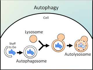
Normally, cells can degrade unwanted proteins.  There are several mechanisms for this, including one called AUTOPHAGY (derived from Greek: auto ("self") + phagein ("to eat")):


Nerdy PowerPoint slide I made. Lysosomes contain acidic digestive enzymes  (like your stomach).
Lysosomes fuse with autophagosomes full of stuff a cell no longer needs and breaks that material down. 

In ALS and the related disease Frontotemporal Lobar Dementia (FTLD), researchers have shown that build up of abnormal TDP-43 is due to a failure of autophagy. 

Scientists from Taiwan are researching ways to turn autophagy back on in ALS and FTLD.  In a recent article, they described how they used chemicals, including the commercially-available drugs tamoxifen (a hormonal drug used to treat breast cancer), rapamycin (used to prevent transplanted organ rejection, and also as a coating for cardiac stents) and carbamazapine (an anti-seizure medication) to stimulate autophagy in mice with FTLD. By doing this, the amount of undigested TDP-43 in nerve cells decreased, and there was less nerve cell death.  This translated into improved strength and memory in the mice. 

But veterinary neurologists are not the only people who should find this heartening: I personally know someone with ALS who is being treated with off-label tamoxifen (along with creatine supplementation).  

I searched www.clinicaltrials.gov and found two trials studying tamoxifen in ALS. One completed accrual in 2008. I could not find any reported results in PubMed or by Googling the terms "amyotrophic lateral sclerosis" and "tamoxifen." I could not find any posted trials of rapamycin for ALS.  

Of interest, protein accumulation due to impaired autophagy may also be relevant in three other neurologic diseases: Huntington's Disease, Parkinson's Disease, and Alzheimer's Dementia. So figuring this out could potentially help a lot of people. 

As "autophagic therapy" (I couldn't make that up) for each of these diseases and ALS is explored, one challenge researchers will face is proving that the treatment is doing what they think its doing (i.e., stimulating autophagy), even if it works to slow or halt disease. I guess that would be a good problem to have, though....

23 February, 2013

Amyloid and Memory

"Whats the last thing that goes through a bug's mind as it hits the windshield of your car?"

The answer to this old joke ("it's a**hole") may turn out only to be true if said bug has defective Orb2, a member of a family of RNA-binding proteins called Cytoplasmic Polyadenylation Element-Binding Proteins (CPEB). If Orb2 is doing its job, the bug might instead have been reflecting on the one that got away at the moment of impact. 

As discussed in an interesting but rather technical article by Mujumdar A, et al., in drosophila (fruit flies) normal Orb2 forms degradation-resistant amyloid-like oligomers at neuronal synapses (in other words, functional amyloid in the connection points between brain nerve cells). These oligomers are essential to the persistence of long-term memory, which in this paper was determined by measuring "courtship suppression memory."  It goes something like this: a male fruit fly, after repeatedly being told to "buzz-off" by an uninterested female fruit fly, eventually learns to stop pestering her and other uninterested females. Normally, this newly-acquired behavior persists for days before waning. Although its tempting to discuss how eerily similar this sounds to a couple of desperate years in middle school, lets focus instead on what happens in fruit flies with mutant Orb2: within 24 hrs, they are back at it, pestering uninterested female fruit flies. A failure of long-term memory. 

Although CPEB is found in mammalian neurons too, and seems to play a role in memory formation, it isn't clear whether this is mediated by functional amyloid formation. As reviewed by J. Richter, stimulation of CPEB-containing nerve cells results in increases in specific neuronal proteins involved in memory persistence. Thus, it appears CPEB's function as a promotor of protein translation, rather than any amyloidogenic properties, may be the basis of its role in mammalian memory. Jury apparently still out, though. 

In the near future, I'll discuss another situation in which amyloid (amyloid-beta) plays a role in memory - in this case, the LOSS of memory - in people with Alzheimer's Dementia.    

21 February, 2013

Pomalidomide and Light Chain Amyloidosis

With the immunomodulatory drug pomalidomide (Pomalyst) recently approved for patients with multiple myeloma who have progressing disease within 60 days of the last of at least 2 prior therapies which had to have included lenalidomide (Revlimid) and bortezomib (Velcade), its an opportune time to review what we know (and don't know) about the drug in light chain (AL) amyloidosis. 

Pomalidomide, like lenalidomide, is a structural analogue of thalidomide. The structures of each are shown here:


source: http://www.readcube.com/articles/10.1186/2162-3619-1-27

The FDA-approved dose of POM in multiple myeloma is 4 mg/day for 21 out of every 28 days.

Investigators at the Mayo Clinic have studied POM (with dexamethasone) in 33 patients with previously treated AL amyloidosis. Patients had had a median of 2 prior therapies, with about half having had autologous stem cell transplant. A few (7) had previously been treated with other immunomodulatory drugs. 

The initial dose of POM in this study was 2 mg/day for 28 consecutive days each cycle (no break). DEX was given at 40 mg/week. POM dosing could be adjusted upward (if no response) and downward (for toxicity). The doses actually received are shown here: 

source: http://bloodjournal.hematologylibrary.org/content/119/23/5397.long

About half of the patients had hematologic responses, with the majority of these being partial. The median duration of response was 19 months. A handful of patients who had hematologic responses also had improvement in organ function, which is about what would be expected (based on prior work involving transplant, bortezomib, and other agents). Some of the responses observed occurred in patients who had had prior lenalidomide. 

A few notes regarding toxicity/adverse events: mild hematologic and gastrointestinal events were common. Peripheral sensory neuropathy (numbness, tingling) was documented in almost all patients - this was generally mild, but was more common than one might expect from, say, lenalidomide (at least in myeloma patients). The authors noted that the cardiac biomarker NT-pro-BNP (a marker of heart failure) sometimes worsened even in the setting of a hematologic response. Similar discordance between light chain measurements and cardiac markers have been noted previously in AL patients treated with immunomodulatory drugs (take a look).  In the present study, a 30% rise in the NT-pro-BNP in the first 3 months of therapy was associated with inferior survival, even though some such patients had concurrent light chain improvement.  

What we know: 
  • POM can (and undoubtedly will) be used as treatment for AL amyloidosis. 
  • Responses can be seen even in patients who have had prior autologous stem cell transplant, bortezomib, or lenalidomide.
What we don't know: 
  • Optimal dose, schedule, and duration of POM in AL amyloidosis (tsk! details!) 
  • Mechanism of NT-pro-BNP increase with POM therapy.
Study Spotlight: 

POM, like lenalidomide and thalidomide, will be studied in combination with other active drugs in patients with AL amyloidosis. The Karmanos Cancer Institute will be coordinating a national trial investigating the combination of POM, bortezomib, and dexamethasone in AL amyloidosis. This is the first trial prospectively studying an immunomodulatory drug combined with a proteasome inhibitor as front-line therapy for AL amyloidosis.  Participating centers will include Boston University, Duke University, the Colorado Blood Cancer Institute (Denver, CO) and Princess Margaret Hospital (Toronto).  

11 February, 2013

"Functional Amyloid"

"Functional amyloid" is a term I first heard about 3 years ago at the XII Amyloidosis Symposium in Rome. There was an oral presentation about the organization of peptide hormones in the secretory granules of the pituitary. Prior to that, I , like most clinicians, only thought about amyloid in the context of disease. It turns out amyloid actually plays an important role in vertebrate and invertebrate biology. 

In bacteria amyloid fibrils are involved in biofilm formation and cell/cell cell/substrate adhesion. Amyloid comprised of curli proteins in E.coli and Salmonella are a well-described example (see figure). Functional amyloid is also produced by Saccharomyces and Candida


source: http://www.mcdb.lsa.umich.edu/labs/chapman/research.php
The oval-shaped things are bacteria; the mesh-like stuff in between them is curli amyloid. 
As in amyloid diseases, the protein fibrils are extracellular; unlike these conditions, the production of amyloid appears to be a highly regulated process with intracellular trafficking, cellular export, fibril nucleation and the rapid polymerization requiring complex interactions between several curlin subunits (CsgA though G).  Its actually pretty interesting. No really, it is.


source: http://www.sciencedaily.com/releases/2008/02/080215121210.htm
A bacterial biofilm. Credit: Janice Haney Carr in Science Daily
In humans, a type of functional amyloid made up of a protein called pmel17 plays a role in the polymerization of the pigment melanin. Smaller melanin precursors (the building blocks from which melanin is ultimately constructed) would be toxic to the cell if they were not sequestered inside melanosomes by pmel17 amyloid. As defined by freemedicaldictionary.com, melanosomes are cigar- or oval-shaped pigment-containing granules within melanocytes. These granules are injected into keratinocytes, providing color:


source: http://www.katzenzeitung.eu/en/Genetics/melanocytes.html
Thus, pmel17 amyloid is critical for the formation of melanin and also cellular protection during this process. Click here for a link to an article discussing this in detail. 

The presentation in Rome I mentioned at the outset of this post discussed yet another functional role for amyloid in humans. Peptide hormones in the pituitary gland are actually stored as amyloid within secretory granules. It is postulated that this structure permits on the one hand, enduring stability in a highly concentrated state, and on the other, a means of controlled release of the hormones in question. Click here to read a publication from the authors of that presentation. 

There are illnesses characterized by abnormal amyloid or amyloid-like protein aggregates inside cells ("inclusion bodies"). An example would be Lewy Bodies in the neurons of patients with Parkinson's Disease. This type of protein accumulation is not functional amyloid, as it is generally damaging to the cell.

One can hope that understanding how cells handle functional intracellular amyloid will eventually provide insights into the pathophysiology of these disease states and perhaps lead to effective treatments. 


27 January, 2013

New Amyloid Fibril Nomenclature Recommendations Published

In May of 2012, at the XIIIth International Symposium on Amyloidosis in Groningen (Netherlands), the Nomenclature Committee of the International Society of Amyloidosis updated amyloid fibril nomenclature guidelines, and expanded the list of recognized amyloid fibril subtypes. The latter is shown here in 3 tables from the article
From Sipe J.D., et al. Amyloid, 2012; 19(4): 167–170

From Sipe J.D., et al. Amyloid, 2012; 19(4): 167–170
From Sipe J.D., et al. Amyloid, 2012; 19(4): 167–170

 I wanted to post the Amyloid Fibril Protein Nomenclature List because it serves as a reference for readers, and also provides context for the broad range of medical topics which will be discussed over time in this blog. Keep this post handy!


26 January, 2013

Suffering from TE

I am suffering from progressive intermittent Theralite Envy (TE), a recently-recognized condition amongst American clinicians who treat a lot of myeloma patients but do not have ready access to the Gambro HCO 1100 dialyzer filter.  The cause of TE can be debated, but my case was definitely exacerbated by recently admitting a myeloma patient with severe acute kidney injury from cast nephropathy which did not improve despite prompt recognition of the problem and initiation of effective systemic myeloma therapy. Several years ago, a consulting physician once assured me that plasmapheresis could cure my TE, but its really only made it worse (see why).

The Gambro HCO ("high cut-off") 1100 dialyzer is distinct for its 15-60 nm pore size, permitting passage of molecules with a MW of up to 45 Daltons (the size of a dimeric lambda light chain).

The Gambro HCO 1100 Dialyzer (*sigh*)
Like Charlie Brown's Little Red Haired Girl, it was a few curves with little else to go on which attracted me to the GHCO1100...

http://www.wikilite.com/wiki/index.php/The_kidney_and_monoclonal_free_light_chains

The figure above models clearance of light chains from the serum with plasma exchange and HCO hemodialysis.

Randomized studies - EuLITE and MYRE - have been ongoing (and ongoing, and ongoing...) in Europe. In both studies, myeloma patients with renal injury who are being treated with bortezomib-based therapy will be randomized to either HCO dialysis with the Gambro filter, or to conventional high flux hemodialysis.  The EuLITE trial was initiated 5 years ago, and results are expected this year.

The technology may have relevance for patients with AL amyloidosis, as well.  The mechanism of renal failure in AL is different, but disease outcome is clearly linked to prompt, deep, sustained reduction in serum light chain levels:

cited in http://www.readcube.com/articles/10.1186/1756-8722-4-47

Since the best novel agent combination regimens for AL amyloidosis typically take over a month to induce maximal light chain suppression, its reasonable to explore HCO dialysis as an adjunctive therapy, either in the initial stages of treatment, or as an ongoing part of care. Dr. Giampaolo Merlini, a world-reknowned amyloidosis specialist from Pavia, Italy is currently conducting a trial using the Gambro filter in AL amyloidosis patients with amyloid heart disease and end-stage kidney injury. In this small study, it may be hard to prove what HCO hemodialysis adds to systemic therapy in terms of light chain reduction, let alone what it adds to the likelihood of inducing an organ response. Dialyzing away the main serum marker used in chemotherapy response assessment may also present a difficulty - particularly when trying to determine the likely benefit of ongoing chemo.

Professor Giampaolo Merlini, Amyloid Guru

At this point, as a sufferer of chronic TE, my prognosis remains guarded: it feels like we have waited an eternity for the results of large and small European trials examining whether use of the Gambro HCO 1100 dialyzer will have real utility in patients with plasma cell diseases. News may come this year.  Negative results would probably relieve me of my longstanding TE, but would also certainly be a disappointment for the myeloma/amyloidosis medical and patient community. The needed prescription then for both patients suffering from light chain mediated kidney and organ disease, as well as clinicians and researchers suffering from TE? Positive results leading to accelerated availability of the filter on this side of the Atlantic. Fingers crossed.

[AUTHOR NOTE: There is no financial relationship of any sort between myself and Gambro, the maker of the product discussed above. As far as I know, I have never even met anyone associated with Gambro. Admitting this, as readers might imagine, is actually aggravating my TE a little bit]

[TE UPDATE, 2/3/13 10:00 PM EST: Have been in contact with Jeffrey R. Shideman, PhD, Director US Clinical Affairs, Gambro this last week regarding the HCO 1100 Dialyzer (AKA Theralite Filter). There is a compassionate use option for myeloma patients with acute light chain-related renal failure in the United States. This requires a little bit of work on the part of the treating physician (IRB approval, for one thing). FDA guidance on access to investigational devices is provided here. Previously discussed Theralite Envy seems somewhat eased by this information.]

20 January, 2013

9 years and counting....

The last U.S.-led randomized study comparing different treatments for newly-diagnosed AL amyloidosis was published in 2004. The one before that was published in 1999. Two randomized studies, 14 years. To put this in some perspective, despite the fact that neither of my two teenage children have expressed any particular interest in conducting large-scale studies in AL amyloidosis, they are only two behind the national medical community during their lifetimes. 

The premature closure of the ECOG-led intergroup E4A08 study, a randomized Phase III trial comparing melphalan and dexamethasone (MD) to MD + bortezomib (MD-Bz) as initial treatment for AL-amyloidosis patients who have not had prior therapy thus represents a missed golden opportunity to advance the field. This was a trial endorsed by SWOG. I served as the SWOG PI for E4A08, and have some insight as to the problems which led to the trial's closure. 

While checking out another amyloidosis-focused site (The Amyloidosis Weekly, edited by a friend and colleague, Bob Orlowski) I found a posted research abstract from the Accrual Working Group (AWG) of the National Cancer Institute's Myeloma Steering Committee which examined barriers to accrual to NCI-sponsored myeloma trials.  Dr. Matthias Weiss, the lead author of the study, provided me with a copy of the poster he presented at the 2012 American Society of Hematology meeting in Atlanta. 


The authors analyzed the results of Survey Monkey surveys returned by 246 researchers and/or support staff affiliated with cooperative oncology research groups like SWOG or ECOG-ACRIN. The surveys asked respondents to rank 10 potential barriers to accrual identified by the AWG in terms of their importance in slowing accrual in large multi-center trials. Predictably, attitudes of community-based respondents differed from those of academic/university-based ones. Dr. Weiss told me he believes barriers 3, 5 and 8 were key to the demise of E4A08, which is plausible, despite the fact that there are NO (ZERO, NADA, ZIPPO) FDA-approved therapeutic agents for initial treatment of AL amyloidosis. I believe a variation of barrier 10 was also a factor: some sites wouldn't allocate the resources needed to open a trial to which they were not likely to accrue more than 1 or 2 patients. Also, shockingly, several reputable investigators at some of the larger SWOG and ECOG centers expressed that they were uncomfortable with the control arm (melphalan plus dexamethasone), and thus wouldn't even open the study. I bet Dr. Jaccard, a leading amyloidosis specialist in France, laughs about that (see why).

Time to look in the mirror and ask ourselves whether we want to remain relevant in the arena of clinical AL amyloidosis research. The next possible standard of care for AL amyloidosis will be determined by our European colleagues who CAN get this study done (and are in the midst of proving it). Shame on us. Lets not allow another 9 years to pass before we get a big up-front AL study done in the United States.