Showing posts with label AL Amyloid. Show all posts

01 December, 2019

#amyloidosisjc 12-1-19: MRD and AL #amyloidosis

Here is a synopsis of the article we will be discussing in the December 1st, 2019 session of #amyloidosisJC, an online journal club focusing on all things amyloidosis.  The synopsis was prepared by two recipients of the Don Brockman ASH 2019 Trainee Travel Grants supported by the Amyloidosis Foundation, Dr Suresh Balasubramanian (@malignantheme, Karmanos Cancer Institute) and Dr Holly Lee (@holly_dldumls, University of Calgary), with the help of Dr Naresh Bumma (@NB191186, Ohio State University), the faculty co-moderator for the session. 

Introduction: 

In this round of #AmyloidosisJC, we will be discussing a clinical paper that discusses the role of minimal (measurable) residual disease (MRD) in the management of AL amyloidosis.


In this retrospective observation study, the authors presented updated results with extended follow up of the AL amyloidosis patients who underwent end of treatment (EOT) MRD assessment by multiparametric flow cytometry (MFC)  described in a previous publication in 2017.


Study objective: 

To establish whether clearance of clonal plasma cells at EOT using sensitive and uniform MFC is associated with improved OS

Patient population: 

Of the original 173 patients with newly diagnosed AL amyloidosis patients (from their 2017 study), 82 patients who had MRD testing at EOT using sensitive and uniform MFC
 were included in this follow up publication.

84% of the patients underwent autologous stem cell transplant as first line therapy (relevance: consider inherent selection bias to include primarily transplant eligible patients who are generally fit and do not have extensive baseline organ involvement
)


Methods: 

For MFC testing, reported sensitivity was 1x10⁻⁴ to 2x10⁻⁵, depending on the number of analyzed events, phenotype and DNA index

A total of 500,000 live cellular events were set as a target per exam (median gated events achieved 489,922, 25–75% IQR 469,765–493,662)

Results:

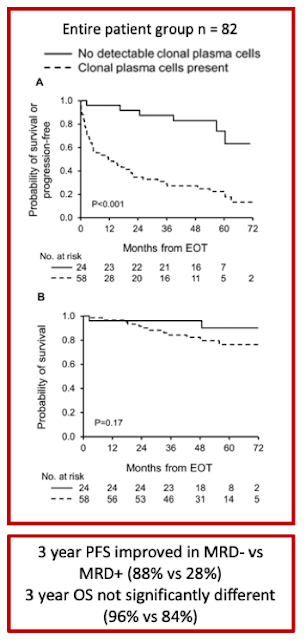
MRD and hematologic response: 29% (24) had negative MRD, and 71% (58) had positive MRD at EOT. 19.5% (16) had CR, 46.3% (38) had VGPR, and 28 (34.1%) had less than VGPR
.


Patient outcomes (median 4.6 year follow up)

Among VGPR patients, MRD- was associated with improved PFS compared to patients with MRD+ (3-year PFS 88% vs. 46%, p=0.003), particularly among patients who achieved a complete response (3-year PFS 100% vs. 33%, p=0.001). In contrast, this difference in PFS advantage in VGPR/MRD- compared to those who achieved VGPR/MRD+ did not reach statistical significance (p=0.14)


OS difference was not seen between MRD- and MRD+ groups (3-year OS 96% vs. 84%, p=0.17)


MRD- compared with MRD+ among deep responders was associated with lower level of involved light chain (involved free light chain, median 1.1 vs. 1.7mg/dL; p=0.02) and higher frequency of renal response (100% vs. 68%; p=0.005)
. When assessing independent organ function, this difference was not evident in cardiac response

Conclusions: 


  • Conclusion 1. Despite the retrospective nature of this study with the inherent selection bias to include primarily transplant eligible patients, this work aims at offering novel, robust surrogate endpoint for the design of clinical trials, as well as for optimizing individual patients’ treatment
  • Conclusion 2. There may be value in bone marrow biopsy/aspirate at end of therapy in AL amyloidosis patients who achieve VGPR/CR. Presence of MRD is associated with reduced PFS in this group of patients.
  • Conclusion 3. The sensitivity of the current standard assay for serum free light chain detection does not discern between MRD positive vs negative among VGPR/CR patients.
  • Conclusion 4. This study is notable for presenting one of the largest patient cohorts for end of treatment MRD assessment and applying uniform flow cytometric techniques.


Discussion:

MRD assessment in AL amyloidosis is not yet standardized. Of interest, a study using next generation flow for MRD assessment found that 5 out of 12 MRD positive cases had very low residual tumor burden (<3x10⁻⁵), which would have not been detected with lower sensitivity assays (Kastritis et al. 2018). This paper assessed 20 AL patients with hemCR. 8 out of 20 patients were MRD(-).   2 out of 8 patients who had ASCT as primary therapy achieved MRD(-) status, versus 6/12 (50%) patients who did not have ASCT as primary therapy (p = 0.264). In two cases aberrant cells were detected at levels between 10−5 and 10−6  Their reported median sensitivity level of next gen flow was 2.3 x 10⁻⁶ (range: 2 × 10−6–3.1 × 10-6)


This is not surprising, as in the myeloma literature, 25% of MRD neg cases by conventional MFC were found to be MRD positive by next gen flow (Flores-Monteroet al. 2017)

We do not have studies that inform us how best to manage/ monitor patients deep hematological response based on their MRD status

  • How does MRD status guide frequency of follow up of patients after therapy?
  • If a patient initially achieves MRD neg CR but then progresses to MRD positive CR on repeat follow up, does this indicate progression/ requirement for treatment?
  • What is the role of transplant in achieving MRD negative status? The impact of daratumumab in achieving MRD negative status? As per our discussion from last week, looking forward to upcoming ISA abstract from @vsanchorawala regarding role of transplant in this topic!
  • Does time to MRD achievement matter and if so, what is the optimal time to assess MRD in AL amyloidosis? Interesting report by Muchtar et al. 2019 in Leukemia journal, looked at not only the depth of response (in this case nadir iFLC <2mg/dL) but also the impact of time to nadir iFLC on patient outcomes. Patients whose nadir iFLC occurred after 12 months from EOT had significantly longer PFS and OS compared to patients who reached nadir before 12 months. This raises the question as to whether there a role for MRD assessment not only at EOT but also further out from EOT.
Obviously, there are many questions related to the utility and usage of MRD testing in AL amyloidosis - looking forward to the journal club where we can explore this further! -J.Zonder,MD

UPDATE 12/1/19 @ 10:19 pm: link to transcript of tonight's twitter discussion CLICK HERE

19 June, 2019

#amyloidosisJC 6/19/19: light chain stabilization and AL #amyloidosis

In this round of #AmyloidosisJC, we will be discussing a basic science paper that describes small molecules that stabilize light chain proteins in vitro. The following summary was written by Gareth Morgan (@wittyremarkhere), the first author. He will lead the discussion for this session of #amyloidosisJC.

Here is a link to the original paper: 

This work was carried out at Scripps Research by the group of Dr. Jeff Kelly. 

Dr. Kelly’s group invented tafamidis, a molecule that stabilizes transthyretin that was recently FDA approved (link: https://www.pfizer.com/news/press-release/press-release-detail/u_s_fda_approves_vyndaqel_and_vyndamax_for_use_in_patients_with_transthyretin_amyloid_cardiomyopathy_a_rare_and_fatal_disease) for treatment of ATTR amyloidosis. 

Background:
Amyloidosis is caused by aggregation of normally-soluble proteins. In inherited diseases such as familial ATTR amyloidosis, aggregation is linked to destabilization of the precursor protein by mutation. In AL amyloidosis this connection is less well understood, because each patient has a unique amyloid-forming antibody light chain, secreted by monoclonal plasma cells. However, several lines of evidence show that unstable light chains are associated with amyloidosis:

2. Blancas-Mejia et al, 2014 https://www.ncbi.nlm.nih.gov/pubmed/24157440

Since stabilization of precursor proteins has been shown to have clinical benefit for ATTR patients, the authors looked for stabilizers of light chains.

Key points from the paper:

1. The authors developed a method to measure light chain stability by high throughput screening. This was important because light chains do not have any known natural ligands that could be modified to make a drug. Instead, screening a large number of molecules was required.

2. From a starting set of 650,000 molecules, 16 molecules in four chemical classes could stabilize light chains when tested in several assays.

3. The paper focuses on one compound, which is a commercially available dye called “coumarin 1”. This molecule becomes fluorescent when it binds to light chains, which makes it useful as a tool for other experiments.

4. The crystal structure of coumarin 1 bound to light chains shows that the small molecule binds between the two variable domains in the dimer, at an interface that is made up of highly conserved residues (Figure D in the image seen at this link: F3.large.jpg). This site is likely to be present in most patient’s involved light chains. However, the molecules do not bind to the normal antibody heavy chain:light chain dimer interface.

5. The authors intend to develop molecules that bind more tightly and more specifically to light chains. These molecules could become drug candidates.

Clinical points for discussion:
1. These molecules are not drugs. There is a lot of work to be done before they can be tested in patients.

2. Stabilization may be most effective in when combined with anti plasma-cell therapies. One potentially promising use would be in maintenance for patients who have a hematological response to therapy but are at risk of relapse. Another would be for patients who are too sick to tolerate cytotoxic drugs.

3. Doxycycline, which has shown some efficacy for AL in Phase 2 trials, does not stabilize light chains in this assay – whatever it’s doing is probably different. https://www.ncbi.nlm.nih.gov/pubmed/28338670

4. Tafamidis is beneficial in ATTR patients. Would a light chain stabilizer have similar properties? We don’t know what the consequences of stabilizing light chains in patients will be. Many individuals with other plasma cell dycrasias (e.g., MGUS, smoldering myeloma, multiple myeloma) tolerate elevated levels of a monoclonal light chain without direct organ damage. However, light chains are cleared by the kidneys and several renal syndromes other than amyloidosis are associated with light chains. Altering light chain metabolism may cause problems in the kidney or elsewhere.

5. if stabilizers alter light chain clearance, they may interfere with free light chain measurements, currently considered key in the management and monitoring of AL amyloidosis.


6. It may be possible to measure the inherent stability of light chains in blood, thereby potentially identifying patients who might benefit from light chains  stabilization by small molecules.

08 May, 2019

#AmyloidosisJC 5/8/19: ASCT in AL patients with impaired renal function

In this installment of #amyloidosisJC we will be discussing outcomes of Autologous Stem Cell Transplant (ASCT) in AL amyloidosis patients who have impaired renal function, written by Dr. M. Hasib Sadiqi and colleagues at the Mayo Clinic's Amyloidosis Program. 

Here is a link to the article: https://www.nature.com/articles/s41409-019-0524-2

For those of you who either do not have access to the full article or didn't have time to read it (or couldn't make the live journal club discussion taking place at 9 pm EST on 5/8/19), here is a synopsis of the paper, including key findings:

  • This is a retrospective review of all patients with AL amyloidosis transplanted at Mayo between May 1999 and September 2017. Of 696 pts, 41 were excluded for various reasons (including ALREADY BEING ON DIALYSIS AT TIME OF ASCT IN 29 PATIENTS).  The remaining 655 pts were divided into "Normal Renal Function" (NRF; eGFR >45 mL/min; n=568) and "Impaired Renal Function" (IRF; eGFR <45 mL/min; n=87)
  • Since this wasn't a randomized prospective study, the groups were predictably not balanced for all clinical characteristics. The NRF grp was less likely to have gotten pre-ASCT chemotherapy (41% vs 53%) and more likely to have been treated with a full dose of melphalan as ASCT conditioning (79% vs 29%). Also, the IRF grp had higher cardiac biomarkers (though no statistical difference in Mayo Cardiac Stage distribution) and more patients with advanced Amyloid Renal Stage (0% renal stage 1, compared with 63% in the NRF grp).  Cardiac and Renal staging have both been covered in previous #amyloidosisJC sessions.
  • 100-day mortality was greater in the IRF grp (14% vs 5%). Risk of progression to ESRD and hemodialysis was also higher in the IRF grp (16% vs 6%). These differences did not have to do with differences in control of the underlying amyloidosis, as the hematologic response rates (88-89%) and complete hematologic response (CR) rates (42-44%) were essentially identical in each grp, though NRF pts who got less than 200 mg/m2 of melphalan had lower CR rates than other patient subsets. There was a higher rate of hospitalizations, a longer duration of hospitalizations, and a higher incidence of culture-confirmed episodes of bacteremia in the IRF group, though causes of death within the first 100 days post-ASCT were not elaborated upon in the article. 
  • Median Overall Survival (140 mos) and Progression Free Survival (49 mos) were not statistically different between the NRF and IRF gaps. 
  • The authors provided a thoughtful discussion regarding the limitations and quirks of the trial. 
    • The period over which patients were treated was long, and eligibility criteria for ASCT evolved over that time. A disproportionate number of pts in the IRF grp were treated in the earlier part of the analyzed period. 
    • It was difficult to determine whether any of the differences in survival were due to an imbalance of cardiac involvement, because cardiac markers (used to assign Cardiac Stage) are affected by renal clearance
    • The definition of "NRF" was not really "N" (that is, a significant number of patients had an eGFR of less than 60 mL/min, though it is not clear exactly how many. The definition of "Renal Stage I" assures us that at least 63% of the NRF pts had an eGFR of at least 50 mL/min). 
Looking forward to a lively discussion at #amyloidosisJC! 

02 November, 2018

#amyloidosisJC 11/4/18: Daratumumab as therapy for AL amyloidosis

We will be discussing this short, sweet, yet practice-changing article demonstrating the significant therapeutic efficacy of daratumumab in a series of patients with AL amyloidosis. Since this publication, there have been numerous other abstracts and a few publications expanding on these findings. We'll discuss them all on Sunday night, 11/4/18, at 8 pm EST.

click HERE for a link to a similar series we just published with colleagues at the Cleveland Clinic, and HERE for an article by Dr Vaishali Sanchorawala (@vsanchorawala) summarizing other data on this topic presented last year at #ASH17

Tweet ya Sunday. I'll be logging in from ALBANIA. Pix of Tirana and from the 13th Congress of the Albanian Association of Hematology to follow. 


21 February, 2016

Leaping back into #amyloidosisJC on Monday 2/29/16 at 9 pm EST: ICD placement for cardiac #amyloidosis

#amyloidosisJC returns at 9 pm EST on Monday February 29th 2016 with a discussion focusing on the role of implanted cardiac defibrillators (ICDs) as a means of improving survival of patients with cardiac amyloidosis.  Thank you to Dr. Naresh Bumma (a Karmanos Cancer Institute hematology-oncology fellow, @NB191186 on Twitter) for his help preparing a summary of the following article from the Mayo Clinic:


Published in the Journal of Cardiovascular Electrophysiology, 2013: 24(7), 793-798.


Background:


Cardiac involvement with systemic amyloidosis by characterized by infiltration and/or deposition of amyloid chains in the myocardium leading to wall thickening and valvular damage.  The presence of cardiac involvement is usually associated with high mortality, in part due to a high risk of fatal arrhythmias. However, the benefit of implantable cardiac defibrillator (ICD) placement in this population remains controversial due to the lack of compelling evidence that it reduces mortality.


Methodology:


Retrospective chart review of all cardiac amyloidosis (CA) patients between 2000 and 2009 seen at a single institution (the Mayo Clinic). All cases of systemic amyloidosis were diagnosed by tissue biopsy and cardiac involvement was established by right ventricular biopsy or echocardiographic findings (left ventricular wall thickness >12 mm in the absence of other etiologies). Patients who underwent ICD implants were identified and characterized. Patients were staged according to the 2004 Mayo staging criteria using troponin T and NTproBNP measurements.


Results:


892 patients were found to have typical features of CA and out of these 53 underwent ICD placement. Of these 53 patients, 33 had AL, 10 had wild-type ATTR, 9 had familial and 1 had AA amyloidosis.
Forty-one patients (77%) underwent ICD placement for primary prevention (18 with unexplained syncope, 9 with left ventricular ejection fraction 35%, 6 with non-sustained VT, and 8 who were considered high risk for other reasons by their treating physican(s)).  Twelve patients (23%) underwent ICD placement for secondary prevention due to sustained ventricular arrhythmia or previous sudden cardiac arrest.


During follow-up (23.25 ± 21.45 months from ICD implantation), 15 patients received at least one appropriate ICD shock, with 12 out of 15 of these occurring in AL patients.


AL amyloidosis subgroup (n=33):


Twelve (36%) underwent successful autologous SCT (including one who underwent orthotopic heart transplantation prior to ASCT) and 21 (64%) were treated with nonmyeloablative chemotherapy.

Median surival was 7.5 months (similar to their historical cohort where median surivival was 10 months, p=0.31)


Familial, AA and wild-type ATTR amyloidosis:


Numbers were insufficient to draw any statistical comparisons


Conclusions:


Despite a high rate of appropriate ICD discharges, there has been no overall survival benefit seen in this cohort with ICD placement for CA compared to cotemporaneous patients without ICD placement.  Possible explanations for these findings are that cardiac deaths due to pump failure (rather than arrhythmias) may account for the observed poor survival in CA patients, or that patient selection for ICD placement needs refinement.

30 November, 2015

#amyloidosisJC 12/1/2015: A staging system for renal outcome and early markers of renal response to chemotherapy in AL amyloidosis.


This is the 5th and final pre-symposium installment of #amyloidosisJC, with the following summary by travel grant awardee and article author Paolo Milani. Dr. Milani is from the esteemed Pavia Amyloidosis group, and is currently spending some time at Mayo Clinic. Needless to say, he'll be bringing a high level of knowledge to this journal club. Starting in January, 2016, #amyloidosisJC will be a monthly online journal club activity.

The article:


Giovanni Palladini1, Ute Hegenbart3, Paolo Milani1, Christoph Kimmich3, Andrea Foli1, Anthony D. Ho3, Marta Vidus Rosini1, Riccardo Albertini2,3, Remigio Moratti5, Giampaolo Merlini1,2,3 and Stefan Schönland3


  1. Amyloidosis Research and Treatment Center, Fondazione Istituto di Ricovero e Cura a Carattere Scientifico (IRCCS) Policlinico San Matteo, Pavia, Italy;
  2. Department of Molecular Medicine, University of Pavia, Pavia, Italy;
  3. Amyloidosis Center, Division of Hematology, Oncology, and Rheumatology, Department of Internal Medicine V, University of Heidelberg, Heidelberg, Germany; and
  4. Clinical Chemistry Laboratory, and 5Scientific Direction, Fondazione IRCCS Policlinico San Matteo, Pavia, Italy

Background:


Immunoglobulin light-chain (AL) amyloidosis is caused by a usually small plasma cell clone synthesizing light chains undergoing conformational changes that lead to their aggregation and deposition in tissues. The kidney is involved in 70% of patients, but little is known on progression or reversibility of renal involvement. Renal involvement results in significant morbidity, and renal failure limits the therapeutic options. Previously, criteria defining renal response and progression were based on the International Society of Amyloidosis (ISA) consensus criteria of 2005 but the clinical significance of each have never been validated. The goal of this study was to identify and validate criteria by assessing the risk of dialysis in 732 consecutive, previously untreated patients with AL amyloidosis with renal involvement evaluated at 2 European referral centers: the Pavia Amyloidosis Research and Treatment Centre and the Heidelberg Amyloidosis Centre.

Methodology:


The databases of the Pavia Amyloidosis Research and Treatment Centre and of the Heidelberg Amyloidosis Centre were systematically searched for subjects with renal involvement diagnosed between 2004 and 2012. Baseline evaluation at each center included a complete physical examination, assessment of amyloid organ involvement, echocardiography, cardiac biomarkers measurement of serum creatinine concentration and of 24-hour urinary protein excretion, serum and urine immunofixation electrophoresis, and quantitation of free light chain (FLC).


The study end point was renal survival, defined as the time from diagnosis to dialysis initiation. The authors assessed the impact of baseline variables and of their changes with therapy on renal survival . The Italian cohort was used as the testing population and the German series as the validating cohort.


Cox models were fitted to compute hazard ratios and 95% confidence intervals for progression to dialysis, identifying baseline variables predicting renal survival. Receiver operating characteristic (ROC) analyses based on progression to dialysis at 2 years identified the thresholds of baseline variables best predicting renal survival.

Results:

Patient Characteristics:

A total of 71 (15%) patients required dialysis in the Italian group and 84 (31%) in the German series.

Patients’ survival from dialysis initiation was not significantly different in the 2 cohorts [median survival was 39 months in the Pavia cohort and 24 months in the Heidelberg cohort (P=0.102)]. Hemodialysis was chosen in most cases, and only 11 patients underwent peritoneal dialysis (9% of the 155 subjects requiring dialysis), 8 in the Italian cohort and 3 in the German cohort.


Factors predicting renal survival:
Renal survival was influenced by proteinuria, eGFR, and, to a lesser extent, by serum albumin. The thresholds best discriminating patients who progressed were 5 g/24 h for urinary protein loss and 50 mL/min per 1.73 m2 for eGFR. Based on these cutoffs, it was possible to design a staging system sharply discriminating 3 groups with significantly different risk of progression to dialysis, with none (renal stage I), 1 (renal stage II), or both (renal stage III) risk factors, respectively (Figure 1A). These results were validated in the Heidelberg cohort (Figure 1B).


Criteria for renal resposne and progression:
For the identification of response and progression criteria, the authors performed a landmark analysis on 472 patients (64%), 301 in the Italian cohort and 171 in the German cohort, who were evaluated for response 6 months after treatment initiation. A ROC analysis based on progression to dialysis at 2 years from the landmark date performed in the testing cohort (Italian series) showed that changes in proteinuria, eGFR, and dFLC (difference between involved [amyloidogenic] and uninvolved free light chain) were able to discriminate subjects progressing to dialysis. The cutoff that identify renal response was a 30% decrease in proteinuria. A 25% decrease in eGFR identified a renal progression. Obtaining a very good partial response (dFLC <40 mg/L) or complete response (negative serum and urine immunofixation and normal FLC ratio) was able to improve renal outcome not only in renal stage II but also in renal stage III patients, indicating that successful treatment can improve renal outcome also in high-risk subjects.  

Finally, they tested the applicability of the novel renal response and progression criteria, related to renal survival, in patients who had response assessment data at an even earlier time point (3 months), 133 in the Italian cohort and 69 in the German cohort, in a 3-month landmark analysis. Given the relatively small number of cases, they performed this analysis in the overall study population of 202 evaluable patients. We found that the proposed response and progression criteria retained their prognostic significance.


Authors’ conclusions:

The progression of renal dysfunction is accurately predicted by baseline proteinuria and eGFR. These 2 variables identify low-risk patients who are unlikely to require dialysis and subjects who are at very high risk of renal failure. Secondly, early changes in eGFR and proteinuria should be used to assess treatment efficacy, in addition to the currently recognized hematologic and cardiac response criteria.

Our comments:

The renal staging system formulated by Palladini and colleagues significantly divided the population of patients with renal AL amayloidosis in low-, intermediate- and high-risk of progression to dialysis. This staging could be a useful tool for the routine patients’ management and could be added in the stratification of patients enrolled in clinical trials.
The identification and validation of renal response and progression criteria that can be assessed at 3 and 6 months is an other important implication of this work. However, it is important to note that the morbidity of renal involvement is not restricted to the minority of patients who develop end stage renal disease.

Finally, we think that it will be important to confirm these data in a prospective setting. Ongoing randomized trials would be the ideal setting.