Showing posts with label Clinical Research. Show all posts

24 March, 2019

#amyloidosisJC 3/27/19: BU Cardiac Staging System for AL Using BNP


This week we will be discussing cardiac staging of AL (light chain) amyloidosis. As mentioned in this post from February 4th, 2018, we are used to the idea of staging AL amyloidosis on the basis of baseline cardiac biomarker measurements. The most widely used system is the European modification of the original 3-stage Mayo 2004 system which uses Troponin T or I and NT-pro-BNP to divide patients into Stages 1, 2, 3a and 3b. The Mayo 2012 system, which incorporates serum free light chain levels, is another 4-stage variation on this theme. Finally, there is a British cardiac staging system for transthyretin (ATTR) amyloidosis which was the subject of a prior #amyloidosisJC discussion. What all of these publications have in common is the use of NT-pro-BNP as a key cardiac biomarker. What all medical centers don't have in common is the ability to order this test without having to send out a blood sample. Many centers use BNP as an alternative. In this installment of #amyloidosisJC, we examine a new 4-stage cardiac staging system for AL amyloidosis devised by researchers at Boston University which incorporates BNP rather than NT-pro-BNP (and we will be lucky enough to have them lead the Twitter discussion!)


Here is a Visual Abstract from the journal Blood summarizing the article:

Visual Abstract for Lilleness B, et al


Key Details of the article we will be reviewing: 
  • A 250-patient derivation cohort from 2016 was used to establish an optimal BNP cut-off to identify cardiac involvement by amyloidosis and also to establish the optimal BNP value to incorporate into a prognostic staging system that corresponded well with the Mayo 2004 system. All the patients in this cohort (except 1) had both BNP and NT-pro-BNP measured. The authors then validated the new staging system with a 1073-patient validation cohort (patients seen between 2004 and 2014, of whom 592 had both BNP and TnI measured at baseline). A Receiver Operating Characteristic (ROC) analysis of 1-yr survival was done to establish a BNP cut-off for BU Cardiac Stage 3b (corresponding to European Cardiac Stage 3b).
  • The presence of cardiac involvement was determined by the following criteria, in order of preference: endomyocardial biopsy or cardiac MRI consistent with cardiac amyloidosis, intraventricular septal end-diastole (IVSd) thickness of at least 12 mm obtained on transthoracic echocardiography without other cause of wall thickening (consistent with established consensus criteria), and IVSd at least 11 mm in men or at least 10 mm in women with no history of hypertension or valvular disease (consistent with current reference ranges, as established by the American Society of Echocardiography).
  • Based on the ECHO/MRI criteria outlined above, 47% of pts in the derivation cohort had cardiac involvement. Only 22 out of the 116 pts identified as having cardiac involvement (19%) had an endomyocardial biopsy confirming cardiac amyloidosis. 
  • In deriving the new cardiac staging system, the authors elected to use the same Troponin I (TnI) cut-off of 0.1 ng/mL used in the Mayo 2004 system, and then determine the best BNP cut-off to use with that. The value they found was 81 pg/mL (𝛋 = 0.854 with Mayo 2004 system). 81 pg/mL also turned out to be an optimal value for predicting the presence of cardiac amyloidosis (except for pts with advanced Chronic Kidney Disease, defined as eGFR <30, in whom the BNP and NT-pro-BNP cut-offs were higher). 
Concordance between he BU and Mayo AL cardiac staging systems
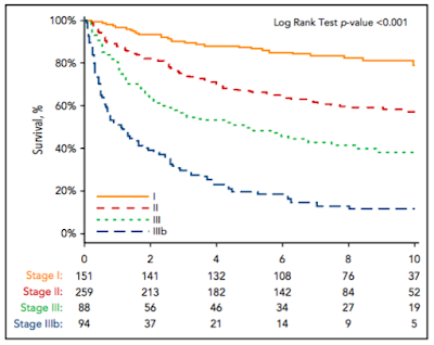
  • Of 592 patients with both BNP and TnI levels measured in the validation cohort, 151 patients (25.5%) were assigned to stage I, 259 patients (43.6%) were assigned to stage II, and 182 patients (30.7%) were assigned to stage III. To create a stage IIIb, a BNP threshold of 700 pg/mL was derived by ROC analysis for survival at 1 yr among the 182 pts with stage III disease (AUC, 0.73; 95% CI, 0.66-0.81). 
  • The median OS from the time of diagnosis was 1.0 year for stage IIIb, 4.3 years for stage III, 9.4 years for stage II, and not reached for stage I patients (Figure 3; HR, 2.6; 95% CI, 1.7-3.9; P , .001). 43.6% of patients with stage IIIb disease died within 6 months.

  • The authors comment on the fact that about half of cardiac stage 3 patients die within the first year, after which time the survival curve for this group flattens out. This is similar to what has been noted previously by the Mayo investigators, and it remains a major challenge in the management of AL amyloidosis. 
  • A BNP-based staging system was hypothesized to be superior to the NT-pro-BNP-based one (since NT-pro-BNP is affected by renal clearance) but this was not confirmed in this work.
Please join us at 9 pm on Wednesday, March 27th, 2019 on Twitter (find us at #amyloidosisJC) for a full and spirited discussion of this article, as well as the other AL cardiac staging papers cited above! 

26 March, 2016

#amyloidosisJC 3/28/16 @ 9 pm EST: Eprodisate for the Treatment of Renal Disease in AA #amyloidosis


This installment of #amyloidosisJC examines the not-positive-enough results of a randomized trial comparing the effects of eprodisate to placebo in AA amyloidosis patients with amyloid-related renal disease. Click HERE for a link to the actual article. I'd like once again to thank Dr. Naresh Bumma (@NB191186) for his assistance preparing this post. 

Dember, Laura M., et al. "Eprodisate for thetreatment of renal disease in AA amyloidosis." New England Journal of Medicine 356.23 (2007): 2349-2360.  

Background: The amyloidoses are a group of diseases characterized by extracellular deposition of fibrils comprised of misfolded proteins and other molecules such as glycosaminoglycans and SAP. End organ damage may result from either the fibrillar deposits or toxic oligomers and/or protofilaments. AA amyloidosis is a rare entity, almost always seen in the setting of an underlying chronic inflammatory condition. The amyloid-forming protein is a proteolytic product of serum amyloid A protein (SAA), an acute phase reactant protein produced by the liver. The kidney is the most commonly affected organ, though the GI tract and other organs can also be involved.

AA Amyloidosis pathophysiology from T. Nakamura (click HERE for link to paper)

Eprodisate (Kiacta), a negatively charged molecule with structural similarities to heparin sulfate, inhibits fibrillogenesis (fibril formation) by interfering with the interaction between SAA and GAGs. The premise of this trial was that if Eprodisate could inhibit fibril formation, organ damage would be prevented. 

Trial Design: Multicenter, randomized, double-blind, placebo-controlled comparison of eprodisate to placebo (1:1 randomization)


13 countries, 27 centers, 261 patients screened and 183 enrolled (7/11/2001-2/14/2003)

Key Inclusion criteria : 
  • Tissue diagnosed AA amyloidosis by histologic demonstration of Congo red staining and birefringence with the use of polarized microscopy and reactivity with anti-AA antibodies by IHC 
  • Kidney involvement (24hr urine protein >1g x 2 OR CrCl <60ml/min x 2)


Key Exclusion criteria: 
  • Kidney disease other than AA amyloidosis, 
  • Severe renal disease: CrCl <20ml/min OR SCr >3, DM
  • Abnormal liver function: LFTs or alk phos >5 xULN, t bili >1.5xULN
  • Diabetes Mellitus
Stratification: 
  • Nephrotic syndrome (Y/N)
  • Treatment center
Patient Allocation: 


Treatment: 800-2400 mg (depending on renal function) eprodisate/placebo in two divided doses each day for 24 months. Alternating office visits and telephone follow-up calls every two months. 

Primary Endpoint: 
  • Composite endpoint of worsening renal function (SCr >2x BL, CrCL <0.5 BL, or progression to dialysis-requiring ESRD) or death

Key Secondary Endpoints:
  • Slope of decline in CrCl over time
  • Change in proteinuria
  • Change in amyloid quantitation in abdominal fat
  • Resolution or development of chronic diarrhea
Noted Patient Characteristics: F>M, ~1/3 pts had "normal" SAA levels, eprodisate group had slightly better baseline SCr (1.1 vs 1.3) but no statistically significant difference in CrCl, ~40% had nephrotic syndrome. The chronic underlying inflammatory condition was usually RA in both groups, but the eprodisate grp had a higher incidence of chronic infection as an alternative condition. 

Results:
  • Primary composite endpoint: 42% reduction in the risk of worsening renal function or death (HR 0.58 [95% CI 0.37 - 0.93; p=0.02]). Teasing this apart: renal function worsened in 27% of eprodisate-treated patients vs 40% placebo-treated pts (p=0.06). There was no difference in mortality (HR 0.95 [95% CI 0.27 - 3.29; p=0.94]).
  • Secondary endpoints: Mean slope (+/- SE) of change in CrCl (in ml/min/1.73m2) was -10.9(+/-5.1) for the eprodisate grp versus -15.6(+/- 4.1) in the placebo grp (p=0.02), though no statistical difference in risk of progressing to ESRD (E: 7 pts, P: 13 pts, HR 0.54 ([95% CI 0.22 - 1.37; p=0.20]). Also, no difference in change in urine protein loss, change in abdominal fat amyloid content, or incidence of developing chronic diarrhea.
  • Safety: Similar rates of AEs and SAEs between grps
Our Analysis: 
  • "Sample size...substantial for the rare disease," but not large enough to be powered to assess differences in lower frequency events like progression to ESRD or death
  • Finite versus indefinite therapy may have mattered
  • PK analysis could have been informative, since more than half of eprodisate pts received modified doses of medication (authors posited that pts with less severe BL renal disease might have been more likely to benefit)
  • Modest benefit may be in part due to mechanism of action: SAA oligomers and protofibril levels not reduced (predictably, since blocking SAA-GAG interaction should only affect mature AA fibril formation). Also, persisting albuminuria itself is nephrotoxic (as explained in an old post about kidney injury in AL amyloidosis) and therapy did not reduce proteinuria.
Have a great Easter (for those who celebrate) and hope a bunch of people can join us this coming Monday!

08 November, 2015

#amyloidosisJC 11/11/15 @ 9 pm EST: targeting the amyloid fibril with anti-SAP therapy


The first two #amyloidosisJC chats were a huge success, and with this installment we hope to build on that. The second journal club, in particular, was fantastic. With Raymond Comenzo (Tufts), Vaishali Sanchorawala (Boston University), and Brendan Weiss & Adam Cohen (both from Penn) participating, it felt like we had the Eastern Conference All-Star team playing. Not to mention over a dozen other engaged participants. This week, with an extremely interesting article on anti-fibrillar therapy being discussed, maybe we can tempt the Western All-Stars into joining...? Mayo? MDACC? Stanford? I'll pester them. Whoever suits up this week, I am certain Drs. Adam Waxman and Brendan Weiss (same Brendan Weiss) will co-moderate a thoughtful and educational discussion.

[special thanks to Dr Waxman for his hard work preparing the original draft of this summary!]

The Article: 

“Therapeutic Clearance of Amyloid by Antibodies to SerumAmyloid P Component.” Richards et. al. NEJM,2015; 373(12):1106-14.


Background:

The systemic amyloidoses are rare diseases characterized by protein misfolding, leading to insoluble amyloid fibril tissue deposition and consequent organ dysfunction. It is the organ deposition, particularly cardiac, that leads to morbidity and mortality. There are over 20 subtypes of systemic amyloidosis. Immunoglobulin light chain (AL) amyloidosis is the most common subtype and is characterized by clonal plasma cell or B-cell expansion and production of misfolded immunoglobulin light chains (LC) that form amyloid and also are directly toxic to organs. Commonly affected tissues and organs include the kidney, liver, heart, and spleen. Currently, the treatment of AL amyloidosis focuses primarily on prevention of light chain production through the use of anti-plasma cell therapy. In other systemic amyloidoses, there are no non-experimental methods to reduce the precursor protein that forms toxic amyloid fibrils. A fundamental problem in the management of the systemic amyloidoses is the development of a treatment to address amyloid that is already deposited in the tissues. A fibril constituent common to all subtypes of amyloidosis is serum amyloid P (SAP). This study explores an early but promising therapy that targets SAP with a monoclonal antibody to stimulate macrophage-mediated clearance of deposited amyloid, which has been previously shown to be effective in a murine model (Bodin, et al. Nature 2010).

The authors (Richards, et al.) performed a phase I clinical trial of a combination of a small molecule CPHPC ((R)-1-[6-[(R)-2-carboxy-pyrrolidin-1-yl]6-oxo-hexanoyl]pyrrolidine-2-carboxylic acid) that has been shown to deplete circulating SAP (serum amyloid P component) followed by administration of a humanized monoclonal anti-SAP antibody that has been shown in preclinical models to stimulate macrophage driven destruction of SAP-containing amyloid deposits. This study describes the first-in-human experience using these agents, including serial assessment of deposited amyloid load in tissues.

Methods:

This is a prospective, single center (National Amyloidosis Center in the United Kingdom), open-label phase I dose escalation study of 16 patients. All patients had biopsy proven systemic amyloidosis; different types of amyloidosis were included. Patients with clinical evidence of cardiac involvement were excluded, as were potentially child bearing women.

CPHPC was administered for 3 days until SAP concentration decreased to below 2.0 mg/L. Patients then received escalating doses of anti-SAP antibody. The last 7 patients received a tailored dose based on the pretreatment SAP load as determined by SAP scintigraphy.

Changes in tissue amyloid content were assessed by whole-body I-SAP scintigraphy at baseline and 42 days post-treatment. Patients were also assessed for retention of tracer at 24 hours by CT. Equilibrium MRI of the heart, liver, and spleen as well as transient elastography (to measure liver stiffness) were performed on days 6, 14, 21, and 42). 42 days was chosen as this was the length of time for SAP to reach equilibrium between the plasma and amyloid components.

Results:

Patient characteristics and response data are shown in Table 1, below. Eight patients had AL amyloidosis, 4 had AFib amyloidosis, 2 had AA amyloidosis, and one had AApoA1. Four of the first 6 patients had small amounts of amyloid in the kidney and spleen. Patient 3 was removed from the study due to poor venous access. Patients 7 – 16 all had moderate to large amounts of amyloid deposition.

Adverse events were mild and included headache and nausea during infusion of CPHPC. Symptoms of transient warmth, flushing, headache, transient changes in heart rate, diarrhea, nausea, and abdominal discomfort during infusion of the anti-SAP antibody were reportedly mitigated with changes in infusion rate. 

There were no changes in serial echocardiography and there were no significant changes in serum troponin T or NT-proBNP concentration.

Rapid depletion of circulating SAP was achieved. Depletion was greater in patients with small or moderate load than in those with a high pre-treatment amyloid load. The half-life of the anti-SAP antibody was found to be around 16 hours in patients with small amyloid load and closer to 4 hours for patients with a large load, consistent with a rapid sequestration.

Correlative studies of immune markers showed that the 9 patients receiving >200 mg of antibody had a transient increase in IL-6, IL-8, CRP, and serum amyloid A protein. Patients receiving >1 mg/kg had a prolonged decrease in complement C3 levels for about a week.

Table 1:


Patient information (amyloid type and response to therapy)

Patients were assessed for amyloid elimination at 42 days after treatment. 6 of 8 patients with liver involvement receiving >200 mg of antibody had a significant decrease in liver stiffness. 5 of these same 8 patients had an overall improvement on their SAP scintigraphy corresponding to decreased amyloid load at 42 days. MRI showed normalization of extracellular volume in 3 patients. Figure 1 shows representative decrease in C3 level and changes in SAP-scintigraphy in patients 8 and 13.

Figure 1:


Response details for two patients 


Author’s Conclusions:

·       Infusion of anti-SAP antibody after CPHPC infusion was safe in patients with systemic amyloidosis with relatively low, transient, infusional side-effects

·       Anti-SAP antibody cleared faster in patients with large hepatic amyloid loads, consistent with rapid binding to their target

·       6/8 patients receiving >200 mg of antibody showed evidence of reduced amyloid load on imaging, even in patients who presumably had continued production of amyloidogenic precursors Including some patients with active AL amyloidosis)

·       Correlative studies of immune markers are consistent with a proposed mechanism of phagocytosis of C3-opsonized complexes by macrophage infiltration as seen in preclinical models

·       Patients with clinically significant cardiac and renal involvement will be included in the next phase of the trial

Our Comments:

·      This small, phase 1 study demonstrates first-in-human data showing a potential decrease in amyloid load among nine out of sixteen enrolled patients with minimal adverse events.

·      Larger confirmatory studies are necessary as is a longer period of follow up to see if decrease in amyloid load at 42 days is a clinically meaningful endpoint

    The optimal dosing and timing of CPHPC and anti-SAP need to be further studied.

·      Patients receiving lower doses of anti-SAP disproportionally had non-AL amyloidosis compared to patients receiving >200 mg and having improvement in amyloid load who mostly (7/9) had AL amyloidosis, which could confound results.

·      Response assessments used non-standard modalities (anti-SAP scintigraphy, liver elastography, extracellular volume by MRI), all of which will need further validation.

·      Delivery of this agent to patients with cardiac disease will need to be done carefully, as the impact of macrophage phagocytosis in the heart on cardiac function is not yet known.

01 November, 2015

#amyloidosisJC 11/4/15 @ 9 pm EST: examining the prognostic and predictive relevance of cytogenetics in AL amyloidosis


CLICK HERE FOR A LINK TO THE FULL TEXT OF THE ARTICLE

Background

Light chain (AL) amyloidosis is a rare plasma cell dyscrasia which causes morbidity and mortality through deposition of toxic amyloid fibrils. Although the prognostic significance of specific cytogenetic aberrations in the plasma cell malignancy multiple myeloma is established, the relevance of cytogenetics in AL amyloidosis is still being defined. Several recent retrospective studies including this study by Bochtler et al., have attempted to characterize the significance of chromosome aberrations in AL amyloidosis. This paper seeks to establish translocation t(11;14), as a predictive marker of poor response to bortezomib. This is important because bortezomib-based regimens (such as Vel/Dex) are often used as front-line treatment for AL amyloidosis. In this disease where patients often have progressive multi-organ damage, selection of an efficacious regimen to rapidly establish disease control is crucial. This paper raises the question of whether cytogenetic studies could guide treatment of AL amyloidosis.

Methods

·      Retrospective, single-center study of 101 consecutive transplant ineligible AL amyloidosis patients who received Vel/Dex as first-line therapy. These patients had high-risk clinical features such as severe cardiac or kidney involvement. Patients with concurrent stage II/III MM or IgM paraprotein were excluded.

·      Outcomes assessed included hematologic response after 3 cycles (by consensus criteria for PR, VGPR), hemEFS (hematologic relapse, hematologic progression, start of a second-line therapy, death) and OS. Early deaths counted as remission failures on intent-to-treat basis.

·       High-risk aberrations were defined as t(4;14), t(14;16) and deletion 17p13.

·      After backward variable selection, validation and calibration of the final multivariable model was done using a CyBorD-treated cohort (32 patients).

Results

Patient Characteristics 

Most patients in Vel/Dex and CyBorD cohorts had cardiac involvement (91% vs 88%) and around half had renal involvement in both cohorts


HemEFS

·      Median hemEFS 4.7 mo with median F/U of 24.0 months and hematologic events in 84/101 patients. The t(11;14)-positive group had inferior hemEFS (median hemEFS 3.4 months vs 8.8 months in t(11;14)-negative group; p=.002). Median hemEFS in high-risk group was 10.3 months vs 3.9 months in patients without high-risk aberrations (P=.15).


Overall Survival (OS)

·      Median OS was 15.7 mo with median F/U time of 24.1 months and 53 observed deaths. t(11;14) predicted for shorter OS with median OS 8.7 months vs 40.7 months in t(11;14)-negative group (P=.05). High risk aberrations conferred favorable prognosis (median OS = NR for high-risk v 10.6 months for absence of high-risk aberrations; P=.04).

Remission Rates

·      95/101 (94%) pt had initial dFLC >50mg/L and thus evaluable for ≥VGPR

·      30 (32%) achieved ≥VGPR after 3 cycles, 24 (25%) attained PR and 21 (22%) did not achieve remission, 20 (21%) had early death before remission assessment. ≥VGPR rates worse in t(11;14)-positive patients compared with t(11;14)-negative patients [14/61 (23%) versus 16/34 (47%) patients, P=.02].  High-risk aberrations had favorable remission rates compared to those without [8/12 (67%) versus 21/80 (26%), P=.008]

Multivariable testing in Vel/Dex Cohort with Clinical and Cytogenetic Factors

·      By Cox regression, t(11;14) and dFLC were the only two statistically significant prognostic markers for both OS and hemEFS. NT-pro-BNP reached statistical signification for OS. Thus t(11;14) is an independent negative risk factor in the Vel/Dex treated cohort. 

    A final model developed: hemEFS, t(11;14), sex, dFLC, NT-proBNP and dose reduction. For OS, t(11;14), dFLC and NT-proBNP. For outcome remission ≥VGPR, t(11;14) and age.

External validation using CyBorD cohort

·      ≥VGPR rate after 3 cycles, 24% (4/17) in t(11;14)-positive compared with 80% (8/10) in t(11;14)-negative patients. Median hemEFS 5.7 months in all 32 patients, 4.0 months in t(11;14)-positive and NR in t(11;14)-negative patients (P=.01). Median OS for both groups NR. Median F/U was 7.5 months. Calibration analysis of final models performed which showed that all trained predictors had a trend for better prediction of the outcome in the CyBorD validation cohort.

Authors’ Conclusions
  • Cytogenetic aberrations are important independent prognostic factors in AL.
  • Bortezomib is less beneficial to patients with t(11;14) 
  • Bortezomib overcomes the poor prognosis of patients with high-risk aberrations.


Our Comments
  • Study was of small sample size and would need confirmation with other studies, prospectively and multi-center ideally
  • A potential criticism is the validation using a CyBord-treated cohort rather than additional Vel/Dex-treated patients. CyBorD cohort followup duration is a lot shorter than the Vel/Dex cohort. Despite this, results seen in the CyBord patients seem consistent with those described for the Vel-Dex cohort
  • Paper suggests that cytogenetics may have predictive as well as prognostic significance.
Special thanks to Sandy Wong (@SandyWong02111) from Tufts University for her efforts preparing this summary. Looking forward to co-moderating this journal club discussion with her. 

20 January, 2013

9 years and counting....

The last U.S.-led randomized study comparing different treatments for newly-diagnosed AL amyloidosis was published in 2004. The one before that was published in 1999. Two randomized studies, 14 years. To put this in some perspective, despite the fact that neither of my two teenage children have expressed any particular interest in conducting large-scale studies in AL amyloidosis, they are only two behind the national medical community during their lifetimes. 

The premature closure of the ECOG-led intergroup E4A08 study, a randomized Phase III trial comparing melphalan and dexamethasone (MD) to MD + bortezomib (MD-Bz) as initial treatment for AL-amyloidosis patients who have not had prior therapy thus represents a missed golden opportunity to advance the field. This was a trial endorsed by SWOG. I served as the SWOG PI for E4A08, and have some insight as to the problems which led to the trial's closure. 

While checking out another amyloidosis-focused site (The Amyloidosis Weekly, edited by a friend and colleague, Bob Orlowski) I found a posted research abstract from the Accrual Working Group (AWG) of the National Cancer Institute's Myeloma Steering Committee which examined barriers to accrual to NCI-sponsored myeloma trials.  Dr. Matthias Weiss, the lead author of the study, provided me with a copy of the poster he presented at the 2012 American Society of Hematology meeting in Atlanta. 


The authors analyzed the results of Survey Monkey surveys returned by 246 researchers and/or support staff affiliated with cooperative oncology research groups like SWOG or ECOG-ACRIN. The surveys asked respondents to rank 10 potential barriers to accrual identified by the AWG in terms of their importance in slowing accrual in large multi-center trials. Predictably, attitudes of community-based respondents differed from those of academic/university-based ones. Dr. Weiss told me he believes barriers 3, 5 and 8 were key to the demise of E4A08, which is plausible, despite the fact that there are NO (ZERO, NADA, ZIPPO) FDA-approved therapeutic agents for initial treatment of AL amyloidosis. I believe a variation of barrier 10 was also a factor: some sites wouldn't allocate the resources needed to open a trial to which they were not likely to accrue more than 1 or 2 patients. Also, shockingly, several reputable investigators at some of the larger SWOG and ECOG centers expressed that they were uncomfortable with the control arm (melphalan plus dexamethasone), and thus wouldn't even open the study. I bet Dr. Jaccard, a leading amyloidosis specialist in France, laughs about that (see why).

Time to look in the mirror and ask ourselves whether we want to remain relevant in the arena of clinical AL amyloidosis research. The next possible standard of care for AL amyloidosis will be determined by our European colleagues who CAN get this study done (and are in the midst of proving it). Shame on us. Lets not allow another 9 years to pass before we get a big up-front AL study done in the United States.