24 March, 2019

#amyloidosisJC 3/27/19: BU Cardiac Staging System for AL Using BNP


This week we will be discussing cardiac staging of AL (light chain) amyloidosis. As mentioned in this post from February 4th, 2018, we are used to the idea of staging AL amyloidosis on the basis of baseline cardiac biomarker measurements. The most widely used system is the European modification of the original 3-stage Mayo 2004 system which uses Troponin T or I and NT-pro-BNP to divide patients into Stages 1, 2, 3a and 3b. The Mayo 2012 system, which incorporates serum free light chain levels, is another 4-stage variation on this theme. Finally, there is a British cardiac staging system for transthyretin (ATTR) amyloidosis which was the subject of a prior #amyloidosisJC discussion. What all of these publications have in common is the use of NT-pro-BNP as a key cardiac biomarker. What all medical centers don't have in common is the ability to order this test without having to send out a blood sample. Many centers use BNP as an alternative. In this installment of #amyloidosisJC, we examine a new 4-stage cardiac staging system for AL amyloidosis devised by researchers at Boston University which incorporates BNP rather than NT-pro-BNP (and we will be lucky enough to have them lead the Twitter discussion!)


Here is a Visual Abstract from the journal Blood summarizing the article:

Visual Abstract for Lilleness B, et al


Key Details of the article we will be reviewing: 
  • A 250-patient derivation cohort from 2016 was used to establish an optimal BNP cut-off to identify cardiac involvement by amyloidosis and also to establish the optimal BNP value to incorporate into a prognostic staging system that corresponded well with the Mayo 2004 system. All the patients in this cohort (except 1) had both BNP and NT-pro-BNP measured. The authors then validated the new staging system with a 1073-patient validation cohort (patients seen between 2004 and 2014, of whom 592 had both BNP and TnI measured at baseline). A Receiver Operating Characteristic (ROC) analysis of 1-yr survival was done to establish a BNP cut-off for BU Cardiac Stage 3b (corresponding to European Cardiac Stage 3b).
  • The presence of cardiac involvement was determined by the following criteria, in order of preference: endomyocardial biopsy or cardiac MRI consistent with cardiac amyloidosis, intraventricular septal end-diastole (IVSd) thickness of at least 12 mm obtained on transthoracic echocardiography without other cause of wall thickening (consistent with established consensus criteria), and IVSd at least 11 mm in men or at least 10 mm in women with no history of hypertension or valvular disease (consistent with current reference ranges, as established by the American Society of Echocardiography).
  • Based on the ECHO/MRI criteria outlined above, 47% of pts in the derivation cohort had cardiac involvement. Only 22 out of the 116 pts identified as having cardiac involvement (19%) had an endomyocardial biopsy confirming cardiac amyloidosis. 
  • In deriving the new cardiac staging system, the authors elected to use the same Troponin I (TnI) cut-off of 0.1 ng/mL used in the Mayo 2004 system, and then determine the best BNP cut-off to use with that. The value they found was 81 pg/mL (𝛋 = 0.854 with Mayo 2004 system). 81 pg/mL also turned out to be an optimal value for predicting the presence of cardiac amyloidosis (except for pts with advanced Chronic Kidney Disease, defined as eGFR <30, in whom the BNP and NT-pro-BNP cut-offs were higher). 
Concordance between he BU and Mayo AL cardiac staging systems
  • Of 592 patients with both BNP and TnI levels measured in the validation cohort, 151 patients (25.5%) were assigned to stage I, 259 patients (43.6%) were assigned to stage II, and 182 patients (30.7%) were assigned to stage III. To create a stage IIIb, a BNP threshold of 700 pg/mL was derived by ROC analysis for survival at 1 yr among the 182 pts with stage III disease (AUC, 0.73; 95% CI, 0.66-0.81). 
  • The median OS from the time of diagnosis was 1.0 year for stage IIIb, 4.3 years for stage III, 9.4 years for stage II, and not reached for stage I patients (Figure 3; HR, 2.6; 95% CI, 1.7-3.9; P , .001). 43.6% of patients with stage IIIb disease died within 6 months.

  • The authors comment on the fact that about half of cardiac stage 3 patients die within the first year, after which time the survival curve for this group flattens out. This is similar to what has been noted previously by the Mayo investigators, and it remains a major challenge in the management of AL amyloidosis. 
  • A BNP-based staging system was hypothesized to be superior to the NT-pro-BNP-based one (since NT-pro-BNP is affected by renal clearance) but this was not confirmed in this work.
Please join us at 9 pm on Wednesday, March 27th, 2019 on Twitter (find us at #amyloidosisJC) for a full and spirited discussion of this article, as well as the other AL cardiac staging papers cited above! 

20 January, 2019

#amyloidosisJC 1/23/19: LECT2 Amyloidosis

#amyloidosisJC resumes this week on a new night: Wednesday night (Jan 23, 2019 at 8 pm EST). 

Short notice and a short but important article describing a series of patients with a relatively recently recognized subtype of amyloidosis: ALect2. 

Here is a link to the full text of the article: 

http://www.bloodjournal.org/content/bloodjournal/123/10/1479.full.pdf

For anyone unable to access the article via that link, email me at zonderj@gmail.com and I can send you the pdf. 

The article we are reviewing this week discusses a series of 130 patients with hepatic #amyloidosis analyzed with tandem mass spectrometry at the Mayo Clinic. 

Key Points:


  • The most common form of hepatic amyloidosis was AL (light chain) subtype. Alect2 also commonly affects the kidneys. 
  • Leukocyte cell-derived chemotaxin 2 associated amyloidosis (ALect2) was next most common (25% of analyzed cases).
  • No definite amyloid-associated LECT2 gene mutation has been identified, but 10 of 10 sequenced patients in another report were homozygous for the G nucleotide in a non-synonymous SNP amongst people of Mexican heritage. 


We'll talk about Alect2 and other forms of hepatic and renal amyloidosis this Wednesday - join us! 

15 November, 2018

#amyloidosisJC 11/18/18: Tafamidis for ATTR amyloidosis!

The following is a GUEST-POST composed by Dr. Sascha Tuchman, a friend and colleague from the University of North Carolina in Chapel Hill, NC. Dr Tuchman will be co-moderating the final pre-ASH 2018 #amyloidosis journal club (#amyloidosisJC) with Untangling Amyloidosis 208 Trainee Travel Grant Awardee Dr. Sam Rubinstein

Dr Tuchman
  
Dr Rubinstein


Background: 

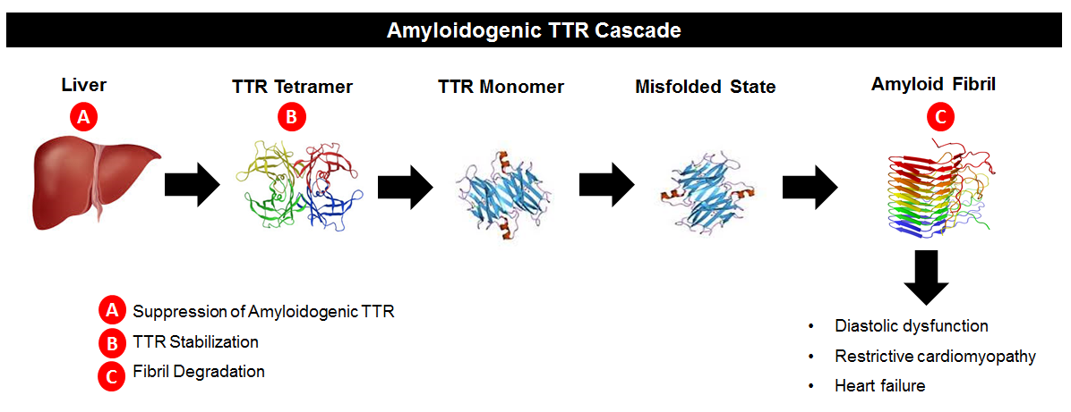
For basic information on transthyretin (TTR) and how it forms amyloidosis, please refer to the background information in recent blog post dated 10/28/18, which summarized the APOLLO study of patisiranin hereditary transthyretin polyneuropathy.  Drugs like patisian work to suppress TTR synthesis by the liver (“A” in the below diagram). Another area of research interest has been in TTR stabilizers. If we refer to the image below, we see that TTR circulates in blood as a tetramer, meaning a single molecule made up of four pieces of TTR joined together. The TTR tetramer dissociates, resulting in single TTR pieces (monomers).  Those monomers can deposit in organs and form amyloid.  The theory behind TTR stabilizers is that making it harder for the tetramers to dissociate means there are fewer TTR monomers available to form amyloidosis (“B” in the below diagram). The “B” approach was first successfully tested using the drug diflunisal in patients with TTR neuropathy (Berk J et al., JAMA 2013).  The current “ATTR-ACT” study examined the oral TTR stabilizer tafamidis in patients with TTR amyloid cardiomyopathy.


Methods: 

Phase 3, international study of 441 patients with biopsy-proven TTR cardiac amyloidosis. Placebo-controlled and double-blinded.  Patients had to have clinical evidence of active heart failure, but less severe than symptomatic at rest (NYHA class <4).  Severe impairment of liver/kidney function or nutritional status precluded participation.  Patients were prohibited from receiving other TTR-directed agents such as diflunisal or doxycycline during study participation. 

Patients received either 80 or 20 mg of tafamidis daily orally or placebo (randomized in a 2:1:2 ratio). Stratified by TTR mutational status (wild-type vs mutated) and baseline NYHA class. Treatment was for 30 months and on completion, patients could opt to continue on to an extension study including open label tafamidis.

The primary endpoint was all-cause mortality followed by cardiovascular-related hospitalizations.  The planned sample size of 400 patients was 90% powered to detect a 30% reduction in mortality and in cardiovascular-related hospitalizations from 2.5 to 1.5 over the 30 months on study.

Results: 

548 patients screened and 441 randomized. 264 patients received tafamidis and 177 placebo. ~24% in both groups had mutated TTR, with a similar frequency of specific TTR mutations in each group. Other baseline factors were similar, including NYHA class, NT-proBNP, and body mass index (BMI).

Both mortality and frequency of cardiovascular-related hospitalizations were improved with tafamidis. Specifically the hazard ratio for all-cause mortality was 0.7 (95% CI 0.51-0.96) and the relative risk for hospitalizations was 0.68 (0.56-0.81).  The mortality benefit appeared after approximately 18 months on study. Benefits were consistent across all subgroups except NYHA class 3 CHF, in which mortality was the same but rate of hospitalizations was higher for tafamidis.
Functional measures of the heart also showed a slower decline with tafamidis than placebo, such as distance walked in 6 minute walk test and patient-reported symptoms.

There were no significant adverse events for tafamidis, and discontinuation from study as a result of adverse events was more common for placebo-treated than tafalidis-treated patients.

Conclusions: 

Tafamidis reduced mortality and cardiovascular-related hospitalization in patients with cardiomyopathy resulting from either wild-type or mutated TTR.  Markers of heart function such as 6 minute walk test and NT-proBNP suggest that tafamidis doesn’t stop the disease, rather it slows its progression.


Comments: 

Tafamidis is clearly a game-changer in the sense that it’s the first medication that will likely be FDA-approved for TTR cardiomyopathy. However, the fact that the disease still progresses indicates that more needs to be done. In particular, combining tafamidis with drugs that perturb other aspects of TTR formation could be promising. (Recall in the diagram above that tafamidis is a “B” drug.  Other drugs such as patisiran and inotersen are “A” drugs.  Combining a B drug with an A drug and/or a C drug may improve the benefit overall to patients with these diseases by further slowing or even reversing damage caused to the heart by TTR.)

EDITOR'S ADDENDUM: looking forward to this week's installment of #amyloidosisJC, at 8 pm EST on Sunday November 18, 2018. Remember to use the #amyloidosisJC hashtag when you log on! Also, hoping to see you in San Diego for the Untangling Amyloidosis 2018 Friday Satellite Symposium. Turns out Dr Tuchman is joining the faculty speaker roster!