01 December, 2019

#amyloidosisjc 12-1-19: MRD and AL #amyloidosis

Here is a synopsis of the article we will be discussing in the December 1st, 2019 session of #amyloidosisJC, an online journal club focusing on all things amyloidosis.  The synopsis was prepared by two recipients of the Don Brockman ASH 2019 Trainee Travel Grants supported by the Amyloidosis Foundation, Dr Suresh Balasubramanian (@malignantheme, Karmanos Cancer Institute) and Dr Holly Lee (@holly_dldumls, University of Calgary), with the help of Dr Naresh Bumma (@NB191186, Ohio State University), the faculty co-moderator for the session. 

Introduction: 

In this round of #AmyloidosisJC, we will be discussing a clinical paper that discusses the role of minimal (measurable) residual disease (MRD) in the management of AL amyloidosis.


In this retrospective observation study, the authors presented updated results with extended follow up of the AL amyloidosis patients who underwent end of treatment (EOT) MRD assessment by multiparametric flow cytometry (MFC)  described in a previous publication in 2017.


Study objective: 

To establish whether clearance of clonal plasma cells at EOT using sensitive and uniform MFC is associated with improved OS

Patient population: 

Of the original 173 patients with newly diagnosed AL amyloidosis patients (from their 2017 study), 82 patients who had MRD testing at EOT using sensitive and uniform MFC
 were included in this follow up publication.

84% of the patients underwent autologous stem cell transplant as first line therapy (relevance: consider inherent selection bias to include primarily transplant eligible patients who are generally fit and do not have extensive baseline organ involvement
)


Methods: 

For MFC testing, reported sensitivity was 1x10⁻⁴ to 2x10⁻⁵, depending on the number of analyzed events, phenotype and DNA index

A total of 500,000 live cellular events were set as a target per exam (median gated events achieved 489,922, 25–75% IQR 469,765–493,662)

Results:

MRD and hematologic response: 29% (24) had negative MRD, and 71% (58) had positive MRD at EOT. 19.5% (16) had CR, 46.3% (38) had VGPR, and 28 (34.1%) had less than VGPR
.


Patient outcomes (median 4.6 year follow up)

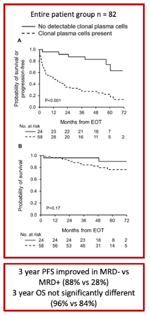
Among VGPR patients, MRD- was associated with improved PFS compared to patients with MRD+ (3-year PFS 88% vs. 46%, p=0.003), particularly among patients who achieved a complete response (3-year PFS 100% vs. 33%, p=0.001). In contrast, this difference in PFS advantage in VGPR/MRD- compared to those who achieved VGPR/MRD+ did not reach statistical significance (p=0.14)


OS difference was not seen between MRD- and MRD+ groups (3-year OS 96% vs. 84%, p=0.17)


MRD- compared with MRD+ among deep responders was associated with lower level of involved light chain (involved free light chain, median 1.1 vs. 1.7mg/dL; p=0.02) and higher frequency of renal response (100% vs. 68%; p=0.005)
. When assessing independent organ function, this difference was not evident in cardiac response

Conclusions: 


  • Conclusion 1. Despite the retrospective nature of this study with the inherent selection bias to include primarily transplant eligible patients, this work aims at offering novel, robust surrogate endpoint for the design of clinical trials, as well as for optimizing individual patients’ treatment
  • Conclusion 2. There may be value in bone marrow biopsy/aspirate at end of therapy in AL amyloidosis patients who achieve VGPR/CR. Presence of MRD is associated with reduced PFS in this group of patients.
  • Conclusion 3. The sensitivity of the current standard assay for serum free light chain detection does not discern between MRD positive vs negative among VGPR/CR patients.
  • Conclusion 4. This study is notable for presenting one of the largest patient cohorts for end of treatment MRD assessment and applying uniform flow cytometric techniques.


Discussion:

MRD assessment in AL amyloidosis is not yet standardized. Of interest, a study using next generation flow for MRD assessment found that 5 out of 12 MRD positive cases had very low residual tumor burden (<3x10⁻⁵), which would have not been detected with lower sensitivity assays (Kastritis et al. 2018). This paper assessed 20 AL patients with hemCR. 8 out of 20 patients were MRD(-).   2 out of 8 patients who had ASCT as primary therapy achieved MRD(-) status, versus 6/12 (50%) patients who did not have ASCT as primary therapy (p = 0.264). In two cases aberrant cells were detected at levels between 10−5 and 10−6  Their reported median sensitivity level of next gen flow was 2.3 x 10⁻⁶ (range: 2 × 10−6–3.1 × 10-6)


This is not surprising, as in the myeloma literature, 25% of MRD neg cases by conventional MFC were found to be MRD positive by next gen flow (Flores-Monteroet al. 2017)

We do not have studies that inform us how best to manage/ monitor patients deep hematological response based on their MRD status

  • How does MRD status guide frequency of follow up of patients after therapy?
  • If a patient initially achieves MRD neg CR but then progresses to MRD positive CR on repeat follow up, does this indicate progression/ requirement for treatment?
  • What is the role of transplant in achieving MRD negative status? The impact of daratumumab in achieving MRD negative status? As per our discussion from last week, looking forward to upcoming ISA abstract from @vsanchorawala regarding role of transplant in this topic!
  • Does time to MRD achievement matter and if so, what is the optimal time to assess MRD in AL amyloidosis? Interesting report by Muchtar et al. 2019 in Leukemia journal, looked at not only the depth of response (in this case nadir iFLC <2mg/dL) but also the impact of time to nadir iFLC on patient outcomes. Patients whose nadir iFLC occurred after 12 months from EOT had significantly longer PFS and OS compared to patients who reached nadir before 12 months. This raises the question as to whether there a role for MRD assessment not only at EOT but also further out from EOT.
Obviously, there are many questions related to the utility and usage of MRD testing in AL amyloidosis - looking forward to the journal club where we can explore this further! -J.Zonder,MD

UPDATE 12/1/19 @ 10:19 pm: link to transcript of tonight's twitter discussion CLICK HERE

19 June, 2019

#amyloidosisJC 6/19/19: light chain stabilization and AL #amyloidosis

In this round of #AmyloidosisJC, we will be discussing a basic science paper that describes small molecules that stabilize light chain proteins in vitro. The following summary was written by Gareth Morgan (@wittyremarkhere), the first author. He will lead the discussion for this session of #amyloidosisJC.

Here is a link to the original paper: 

This work was carried out at Scripps Research by the group of Dr. Jeff Kelly. 

Dr. Kelly’s group invented tafamidis, a molecule that stabilizes transthyretin that was recently FDA approved (link: https://www.pfizer.com/news/press-release/press-release-detail/u_s_fda_approves_vyndaqel_and_vyndamax_for_use_in_patients_with_transthyretin_amyloid_cardiomyopathy_a_rare_and_fatal_disease) for treatment of ATTR amyloidosis. 

Background:
Amyloidosis is caused by aggregation of normally-soluble proteins. In inherited diseases such as familial ATTR amyloidosis, aggregation is linked to destabilization of the precursor protein by mutation. In AL amyloidosis this connection is less well understood, because each patient has a unique amyloid-forming antibody light chain, secreted by monoclonal plasma cells. However, several lines of evidence show that unstable light chains are associated with amyloidosis:

2. Blancas-Mejia et al, 2014 https://www.ncbi.nlm.nih.gov/pubmed/24157440

Since stabilization of precursor proteins has been shown to have clinical benefit for ATTR patients, the authors looked for stabilizers of light chains.

Key points from the paper:

1. The authors developed a method to measure light chain stability by high throughput screening. This was important because light chains do not have any known natural ligands that could be modified to make a drug. Instead, screening a large number of molecules was required.

2. From a starting set of 650,000 molecules, 16 molecules in four chemical classes could stabilize light chains when tested in several assays.

3. The paper focuses on one compound, which is a commercially available dye called “coumarin 1”. This molecule becomes fluorescent when it binds to light chains, which makes it useful as a tool for other experiments.

4. The crystal structure of coumarin 1 bound to light chains shows that the small molecule binds between the two variable domains in the dimer, at an interface that is made up of highly conserved residues (Figure D in the image seen at this link: F3.large.jpg). This site is likely to be present in most patient’s involved light chains. However, the molecules do not bind to the normal antibody heavy chain:light chain dimer interface.

5. The authors intend to develop molecules that bind more tightly and more specifically to light chains. These molecules could become drug candidates.

Clinical points for discussion:
1. These molecules are not drugs. There is a lot of work to be done before they can be tested in patients.

2. Stabilization may be most effective in when combined with anti plasma-cell therapies. One potentially promising use would be in maintenance for patients who have a hematological response to therapy but are at risk of relapse. Another would be for patients who are too sick to tolerate cytotoxic drugs.

3. Doxycycline, which has shown some efficacy for AL in Phase 2 trials, does not stabilize light chains in this assay – whatever it’s doing is probably different. https://www.ncbi.nlm.nih.gov/pubmed/28338670

4. Tafamidis is beneficial in ATTR patients. Would a light chain stabilizer have similar properties? We don’t know what the consequences of stabilizing light chains in patients will be. Many individuals with other plasma cell dycrasias (e.g., MGUS, smoldering myeloma, multiple myeloma) tolerate elevated levels of a monoclonal light chain without direct organ damage. However, light chains are cleared by the kidneys and several renal syndromes other than amyloidosis are associated with light chains. Altering light chain metabolism may cause problems in the kidney or elsewhere.

5. if stabilizers alter light chain clearance, they may interfere with free light chain measurements, currently considered key in the management and monitoring of AL amyloidosis.


6. It may be possible to measure the inherent stability of light chains in blood, thereby potentially identifying patients who might benefit from light chains  stabilization by small molecules.

08 May, 2019

#AmyloidosisJC 5/8/19: ASCT in AL patients with impaired renal function

In this installment of #amyloidosisJC we will be discussing outcomes of Autologous Stem Cell Transplant (ASCT) in AL amyloidosis patients who have impaired renal function, written by Dr. M. Hasib Sadiqi and colleagues at the Mayo Clinic's Amyloidosis Program. 

Here is a link to the article: https://www.nature.com/articles/s41409-019-0524-2

For those of you who either do not have access to the full article or didn't have time to read it (or couldn't make the live journal club discussion taking place at 9 pm EST on 5/8/19), here is a synopsis of the paper, including key findings:

  • This is a retrospective review of all patients with AL amyloidosis transplanted at Mayo between May 1999 and September 2017. Of 696 pts, 41 were excluded for various reasons (including ALREADY BEING ON DIALYSIS AT TIME OF ASCT IN 29 PATIENTS).  The remaining 655 pts were divided into "Normal Renal Function" (NRF; eGFR >45 mL/min; n=568) and "Impaired Renal Function" (IRF; eGFR <45 mL/min; n=87)
  • Since this wasn't a randomized prospective study, the groups were predictably not balanced for all clinical characteristics. The NRF grp was less likely to have gotten pre-ASCT chemotherapy (41% vs 53%) and more likely to have been treated with a full dose of melphalan as ASCT conditioning (79% vs 29%). Also, the IRF grp had higher cardiac biomarkers (though no statistical difference in Mayo Cardiac Stage distribution) and more patients with advanced Amyloid Renal Stage (0% renal stage 1, compared with 63% in the NRF grp).  Cardiac and Renal staging have both been covered in previous #amyloidosisJC sessions.
  • 100-day mortality was greater in the IRF grp (14% vs 5%). Risk of progression to ESRD and hemodialysis was also higher in the IRF grp (16% vs 6%). These differences did not have to do with differences in control of the underlying amyloidosis, as the hematologic response rates (88-89%) and complete hematologic response (CR) rates (42-44%) were essentially identical in each grp, though NRF pts who got less than 200 mg/m2 of melphalan had lower CR rates than other patient subsets. There was a higher rate of hospitalizations, a longer duration of hospitalizations, and a higher incidence of culture-confirmed episodes of bacteremia in the IRF group, though causes of death within the first 100 days post-ASCT were not elaborated upon in the article. 
  • Median Overall Survival (140 mos) and Progression Free Survival (49 mos) were not statistically different between the NRF and IRF gaps. 
  • The authors provided a thoughtful discussion regarding the limitations and quirks of the trial. 
    • The period over which patients were treated was long, and eligibility criteria for ASCT evolved over that time. A disproportionate number of pts in the IRF grp were treated in the earlier part of the analyzed period. 
    • It was difficult to determine whether any of the differences in survival were due to an imbalance of cardiac involvement, because cardiac markers (used to assign Cardiac Stage) are affected by renal clearance
    • The definition of "NRF" was not really "N" (that is, a significant number of patients had an eGFR of less than 60 mL/min, though it is not clear exactly how many. The definition of "Renal Stage I" assures us that at least 63% of the NRF pts had an eGFR of at least 50 mL/min). 
Looking forward to a lively discussion at #amyloidosisJC! 

24 March, 2019

#amyloidosisJC 3/27/19: BU Cardiac Staging System for AL Using BNP


This week we will be discussing cardiac staging of AL (light chain) amyloidosis. As mentioned in this post from February 4th, 2018, we are used to the idea of staging AL amyloidosis on the basis of baseline cardiac biomarker measurements. The most widely used system is the European modification of the original 3-stage Mayo 2004 system which uses Troponin T or I and NT-pro-BNP to divide patients into Stages 1, 2, 3a and 3b. The Mayo 2012 system, which incorporates serum free light chain levels, is another 4-stage variation on this theme. Finally, there is a British cardiac staging system for transthyretin (ATTR) amyloidosis which was the subject of a prior #amyloidosisJC discussion. What all of these publications have in common is the use of NT-pro-BNP as a key cardiac biomarker. What all medical centers don't have in common is the ability to order this test without having to send out a blood sample. Many centers use BNP as an alternative. In this installment of #amyloidosisJC, we examine a new 4-stage cardiac staging system for AL amyloidosis devised by researchers at Boston University which incorporates BNP rather than NT-pro-BNP (and we will be lucky enough to have them lead the Twitter discussion!)


Here is a Visual Abstract from the journal Blood summarizing the article:

Visual Abstract for Lilleness B, et al


Key Details of the article we will be reviewing: 
  • A 250-patient derivation cohort from 2016 was used to establish an optimal BNP cut-off to identify cardiac involvement by amyloidosis and also to establish the optimal BNP value to incorporate into a prognostic staging system that corresponded well with the Mayo 2004 system. All the patients in this cohort (except 1) had both BNP and NT-pro-BNP measured. The authors then validated the new staging system with a 1073-patient validation cohort (patients seen between 2004 and 2014, of whom 592 had both BNP and TnI measured at baseline). A Receiver Operating Characteristic (ROC) analysis of 1-yr survival was done to establish a BNP cut-off for BU Cardiac Stage 3b (corresponding to European Cardiac Stage 3b).
  • The presence of cardiac involvement was determined by the following criteria, in order of preference: endomyocardial biopsy or cardiac MRI consistent with cardiac amyloidosis, intraventricular septal end-diastole (IVSd) thickness of at least 12 mm obtained on transthoracic echocardiography without other cause of wall thickening (consistent with established consensus criteria), and IVSd at least 11 mm in men or at least 10 mm in women with no history of hypertension or valvular disease (consistent with current reference ranges, as established by the American Society of Echocardiography).
  • Based on the ECHO/MRI criteria outlined above, 47% of pts in the derivation cohort had cardiac involvement. Only 22 out of the 116 pts identified as having cardiac involvement (19%) had an endomyocardial biopsy confirming cardiac amyloidosis. 
  • In deriving the new cardiac staging system, the authors elected to use the same Troponin I (TnI) cut-off of 0.1 ng/mL used in the Mayo 2004 system, and then determine the best BNP cut-off to use with that. The value they found was 81 pg/mL (𝛋 = 0.854 with Mayo 2004 system). 81 pg/mL also turned out to be an optimal value for predicting the presence of cardiac amyloidosis (except for pts with advanced Chronic Kidney Disease, defined as eGFR <30, in whom the BNP and NT-pro-BNP cut-offs were higher). 
Concordance between he BU and Mayo AL cardiac staging systems
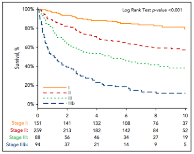
  • Of 592 patients with both BNP and TnI levels measured in the validation cohort, 151 patients (25.5%) were assigned to stage I, 259 patients (43.6%) were assigned to stage II, and 182 patients (30.7%) were assigned to stage III. To create a stage IIIb, a BNP threshold of 700 pg/mL was derived by ROC analysis for survival at 1 yr among the 182 pts with stage III disease (AUC, 0.73; 95% CI, 0.66-0.81). 
  • The median OS from the time of diagnosis was 1.0 year for stage IIIb, 4.3 years for stage III, 9.4 years for stage II, and not reached for stage I patients (Figure 3; HR, 2.6; 95% CI, 1.7-3.9; P , .001). 43.6% of patients with stage IIIb disease died within 6 months.

  • The authors comment on the fact that about half of cardiac stage 3 patients die within the first year, after which time the survival curve for this group flattens out. This is similar to what has been noted previously by the Mayo investigators, and it remains a major challenge in the management of AL amyloidosis. 
  • A BNP-based staging system was hypothesized to be superior to the NT-pro-BNP-based one (since NT-pro-BNP is affected by renal clearance) but this was not confirmed in this work.
Please join us at 9 pm on Wednesday, March 27th, 2019 on Twitter (find us at #amyloidosisJC) for a full and spirited discussion of this article, as well as the other AL cardiac staging papers cited above! 

20 January, 2019

#amyloidosisJC 1/23/19: LECT2 Amyloidosis

#amyloidosisJC resumes this week on a new night: Wednesday night (Jan 23, 2019 at 8 pm EST). 

Short notice and a short but important article describing a series of patients with a relatively recently recognized subtype of amyloidosis: ALect2. 

Here is a link to the full text of the article: 

http://www.bloodjournal.org/content/bloodjournal/123/10/1479.full.pdf

For anyone unable to access the article via that link, email me at zonderj@gmail.com and I can send you the pdf. 

The article we are reviewing this week discusses a series of 130 patients with hepatic #amyloidosis analyzed with tandem mass spectrometry at the Mayo Clinic. 

Key Points:


  • The most common form of hepatic amyloidosis was AL (light chain) subtype. Alect2 also commonly affects the kidneys. 
  • Leukocyte cell-derived chemotaxin 2 associated amyloidosis (ALect2) was next most common (25% of analyzed cases).
  • No definite amyloid-associated LECT2 gene mutation has been identified, but 10 of 10 sequenced patients in another report were homozygous for the G nucleotide in a non-synonymous SNP amongst people of Mexican heritage. 


We'll talk about Alect2 and other forms of hepatic and renal amyloidosis this Wednesday - join us!